2OT0

Fructose-1,6-bisphosphate aldolase from rabbit muscle in complex with a C-terminal peptide of Wiskott-Aldrich syndrome protein


Experimental Data Snapshot

  • Method: X-RAY DIFFRACTION
  • Resolution: 2.05 Å
  • R-Value Free: 0.200 
  • R-Value Work: 0.152 

wwPDB Validation   3D Report Full Report


This is version 1.3 of the entry. See complete history


Literature

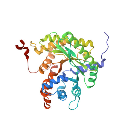
A hydrophobic pocket in the active site of glycolytic aldolase mediates interactions with wiskott-Aldrich syndrome protein.

St-Jean, M.Izard, T.Sygusch, J.

(2007) J Biol Chem 282: 14309-14315

  • DOI: https://doi.org/10.1074/jbc.M611505200
  • Primary Citation of Related Structures:  
    2OT0, 2OT1

  • PubMed Abstract: 

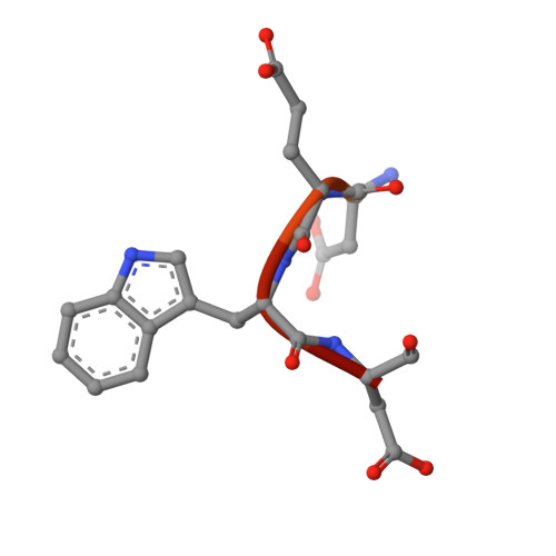
    Aldolase plays essential catalytic roles in glycolysis and gluconeogenesis. However, aldolase is a highly abundant protein that is remarkably promiscuous in its interactions with other cellular proteins. In particular, aldolase binds to highly acidic amino acid sequences, including the C terminus of the Wiskott-Aldrich syndrome protein, an actin nucleation-promoting factor. Here we report the crystal structure of tetrameric rabbit muscle aldolase in complex with a C-terminal peptide of Wiskott-Aldrich syndrome protein. Aldolase recognizes a short, four-residue DEWD motif (residues 498-501), which adopts a loose hairpin turn that folds around the central aromatic residue, enabling its tryptophan side chain to fit into a hydrophobic pocket in the active site of aldolase. The flanking acidic residues in this binding motif provide further interactions with conserved aldolase active site residues Arg-42 and Arg-303, aligning their side chains and forming the sides of the hydrophobic pocket. The binding of Wiskott-Aldrich syndrome protein to aldolase precludes intramolecular interactions of its C terminus with its active site and is competitive with substrate as well as with binding by actin and cortactin. Finally, based on this structure, a novel naphthol phosphate-based inhibitor of aldolase was identified, and its structure in complex with aldolase demonstrated mimicry of the Wiskott-Aldrich syndrome protein-aldolase interaction. The data support a model whereby aldolase exists in distinct forms that regulate glycolysis or actin dynamics.


  • Organizational Affiliation

    Department of Biochemistry, University of Montreal, Montreal, Quebec, Canada.


Macromolecules
Find similar proteins by:  (by identity cutoff)  |  3D Structure
Entity ID: 1
MoleculeChains Sequence LengthOrganismDetailsImage
Fructose-bisphosphate aldolase A
A, B, C, D
363Oryctolagus cuniculusMutation(s): 0 
Gene Names: ALDOA
EC: 4.1.2.13
UniProt
Find proteins for P00883 (Oryctolagus cuniculus)
Explore P00883 
Go to UniProtKB:  P00883
Entity Groups  
Sequence Clusters30% Identity50% Identity70% Identity90% Identity95% Identity100% Identity
UniProt GroupP00883
Sequence Annotations
Expand
  • Reference Sequence

Find similar proteins by:  Sequence   |   3D Structure  

Entity ID: 2
MoleculeChains Sequence LengthOrganismDetailsImage
Wiskott-Aldrich syndrome protein C-terminal peptide
E, F, G, H
15N/AMutation(s): 0 
UniProt & NIH Common Fund Data Resources
Find proteins for P42768 (Homo sapiens)
Explore P42768 
Go to UniProtKB:  P42768
PHAROS:  P42768
GTEx:  ENSG00000015285 
Entity Groups  
Sequence Clusters30% Identity50% Identity70% Identity90% Identity95% Identity100% Identity
UniProt GroupP42768
Sequence Annotations
Expand
  • Reference Sequence
Experimental Data & Validation

Experimental Data

  • Method: X-RAY DIFFRACTION
  • Resolution: 2.05 Å
  • R-Value Free: 0.200 
  • R-Value Work: 0.152 
  • Space Group: P 1 21 1
Unit Cell:
Length ( Å )Angle ( ˚ )
a = 84.084α = 90
b = 56.705β = 97.78
c = 156.188γ = 90
Software Package:
Software NamePurpose
CBASSdata collection
CNSrefinement
HKL-2000data reduction
HKL-2000data scaling
CNSphasing

Structure Validation

View Full Validation Report


Entry History 

Deposition Data

Revision History  (Full details and data files)

  • Version 1.0: 2007-02-27
    Type: Initial release
  • Version 1.1: 2008-05-01
    Changes: Version format compliance
  • Version 1.2: 2011-07-13
    Changes: Version format compliance
  • Version 1.3: 2023-08-30
    Changes: Data collection, Database references, Refinement description