2OKX

Crystal structure of GH78 family rhamnosidase of Bacillus SP. GL1 AT 1.9 A


Experimental Data Snapshot

  • Method: X-RAY DIFFRACTION
  • Resolution: 1.90 Å
  • R-Value Free: 0.214 
  • R-Value Work: 0.182 
  • R-Value Observed: 0.182 

wwPDB Validation   3D Report Full Report


This is version 1.4 of the entry. See complete history


Literature

Crystal Structure of Glycoside Hydrolase Family 78 alpha-L-Rhamnosidase from Bacillus sp. GL1

Cui, Z.Maruyama, Y.Mikami, B.Hashimoto, W.Murata, K.

(2007) J Mol Biol 374: 384-398

  • DOI: https://doi.org/10.1016/j.jmb.2007.09.003
  • Primary Citation of Related Structures:  
    2OKX

  • PubMed Abstract: 

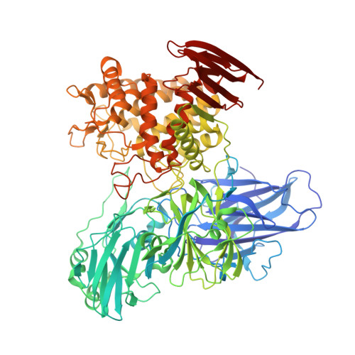
    alpha-L-Rhamnosidase (EC 3.2.1.40) catalyzes the hydrolytic release of rhamnose from polysaccharides and glycosides. Bacillus sp. GL1 alpha-L-rhamnosidase (RhaB), a member of glycoside hydrolase (GH) family 78, is responsible for degrading the bacterial biofilm gellan, and also functions as a debittering agent for citrus fruit in the food and beverage industries through the release of rhamnose from plant glycoside, naringin. The X-ray crystal structure of RhaB was determined by single-wavelength anomalous diffraction using a selenomethionine derivative and refined at 1.9 A resolution with a final R-factor of 18.2%. As is seen in the homodimeric form of the active enzyme, the structure of RhaB in crystal packing is a homodimer containing 1908 amino acids (residues 3-956), 43 glycerol molecules, four calcium ions, and 1755 water molecules. The overall structure consists of five domains, four of which are beta-sandwich structures designated as domains N, D1, D2, and C, and an (alpha/alpha)(6)-barrel structure designated as domain A. Structural comparison by DALI showed that RhaB shares its highest level of structural similarity with chitobiose phosphorylase (Z score of 25.3). The structure of RhaB in complex with the reaction product rhamnose (inhibitor constant, K(i)=1.8 mM) was also determined and refined at 2.1 A with a final R-factor of 19.5%. Rhamnose is bound to the deep cleft of the (alpha/alpha)(6)-barrel domain, as is seen in the clan-L GHs. Several negatively charged residues, such as Asp567, Glu572, Asp579, and Glu841, conserved in GH family 78 enzymes, interact with rhamnose, and RhaB mutants of these residues have drastically reduced enzyme activity, indicating that the residues are crucial for enzyme catalysis and/or substrate binding. To our knowledge, this is the first report on the determination of the crystal structure of alpha-L-rhamnosidase and identification of its clan-L (alpha/alpha)(6)-barrel as a catalytic domain.


  • Organizational Affiliation

    Division of Food Science and Biotechnology, Graduate School of Agriculture, Kyoto University, Uji, Kyoto 611-0011, Japan.


Macromolecules
Find similar proteins by:  (by identity cutoff)  |  3D Structure
Entity ID: 1
MoleculeChains Sequence LengthOrganismDetailsImage
Rhamnosidase B
A, B
956Bacillus sp. GL1Mutation(s): 0 
Gene Names: rhaB
EC: 3.2.1.40
UniProt
Find proteins for Q93RE7 (Bacillus sp. (strain GL1))
Explore Q93RE7 
Go to UniProtKB:  Q93RE7
Entity Groups  
Sequence Clusters30% Identity50% Identity70% Identity90% Identity95% Identity100% Identity
UniProt GroupQ93RE7
Sequence Annotations
Expand
  • Reference Sequence
Small Molecules
Ligands 2 Unique
IDChains Name / Formula / InChI Key2D Diagram3D Interactions
GOL
Query on GOL

Download Ideal Coordinates CCD File 
BA [auth B]
CA [auth B]
DA [auth B]
E [auth A]
EA [auth B]
BA [auth B],
CA [auth B],
DA [auth B],
E [auth A],
EA [auth B],
F [auth A],
FA [auth B],
G [auth A],
GA [auth B],
H [auth A],
HA [auth B],
I [auth A],
IA [auth B],
J [auth A],
JA [auth B],
K [auth A],
KA [auth B],
L [auth A],
LA [auth B],
M [auth A],
MA [auth B],
N [auth A],
NA [auth B],
O [auth A],
OA [auth B],
P [auth A],
PA [auth B],
Q [auth A],
QA [auth B],
R [auth A],
RA [auth B],
S [auth A],
SA [auth B],
T [auth A],
TA [auth B],
U [auth A],
UA [auth B],
V [auth A],
VA [auth B],
W [auth A],
WA [auth B],
X [auth A],
Y [auth A]
GLYCEROL
C3 H8 O3
PEDCQBHIVMGVHV-UHFFFAOYSA-N
CA
Query on CA

Download Ideal Coordinates CCD File 
AA [auth B],
C [auth A],
D [auth A],
Z [auth B]
CALCIUM ION
Ca
BHPQYMZQTOCNFJ-UHFFFAOYSA-N
Experimental Data & Validation

Experimental Data

  • Method: X-RAY DIFFRACTION
  • Resolution: 1.90 Å
  • R-Value Free: 0.214 
  • R-Value Work: 0.182 
  • R-Value Observed: 0.182 
  • Space Group: P 21 21 21
Unit Cell:
Length ( Å )Angle ( ˚ )
a = 97.744α = 90
b = 119.985β = 90
c = 207.352γ = 90
Software Package:
Software NamePurpose
DENZOdata reduction
SCALEPACKdata scaling
SHELXphasing
RESOLVEphasing
CNSrefinement
PDB_EXTRACTdata extraction
HKL-2000data scaling
CNSphasing

Structure Validation

View Full Validation Report


Entry History 

Deposition Data

Revision History  (Full details and data files)

  • Version 1.0: 2007-11-13
    Type: Initial release
  • Version 1.1: 2011-07-13
    Changes: Derived calculations, Source and taxonomy, Version format compliance
  • Version 1.2: 2017-10-18
    Changes: Refinement description
  • Version 1.3: 2023-12-27
    Changes: Data collection, Database references, Derived calculations
  • Version 1.4: 2024-04-03
    Changes: Refinement description