2O0T

The three dimensional structure of diaminopimelate decarboxylase from Mycobacterium tuberculosis reveals a tetrameric enzyme organisation


Experimental Data Snapshot

  • Method: X-RAY DIFFRACTION
  • Resolution: 2.33 Å
  • R-Value Free: 0.241 
  • R-Value Work: 0.191 
  • R-Value Observed: 0.192 

wwPDB Validation   3D Report Full Report


This is version 1.4 of the entry. See complete history


Literature

The three-dimensional structure of diaminopimelate decarboxylase from Mycobacterium tuberculosis reveals a tetrameric enzyme organisation.

Weyand, S.Kefala, G.Svergun, D.I.Weiss, M.S.

(2009) J Struct Funct Genomics 10: 209-217

  • DOI: https://doi.org/10.1007/s10969-009-9065-z
  • Primary Citation of Related Structures:  
    2O0T

  • PubMed Abstract: 

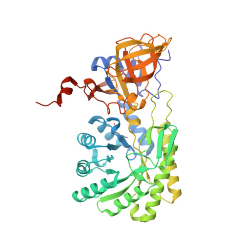
    The three-dimensional structure of the enzyme diaminopimelate decarboxylase from Mycobacterium tuberculosis has been determined in a new crystal form and refined to a resolution of 2.33 A. The monoclinic crystals contain one tetramer exhibiting D(2)-symmetry in the asymmetric unit. The tetramer exhibits a donut-like structure with a hollow interior. All four active sites are accessible only from the interior of the tetrameric assembly. Small-angle X-ray scattering indicates that in solution the predominant oligomeric species of the protein is a dimer, but also that higher oligomers exist at higher protein concentrations. The observed scattering data are best explained by assuming a dimer-tetramer equilibrium with about 7% tetramers present in solution. Consequently, at the elevated protein concentrations in the crowded environment inside the cell the observed tetramer may constitute the biologically relevant functional unit of the enzyme.


  • Organizational Affiliation

    EMBL Hamburg Outstation, c/o DESY, Hamburg, Germany.


Macromolecules
Find similar proteins by:  (by identity cutoff)  |  3D Structure
Entity ID: 1
MoleculeChains Sequence LengthOrganismDetailsImage
Diaminopimelate decarboxylase
A, B, C, D
467Mycobacterium tuberculosis H37RvMutation(s): 0 
Gene Names: lysA
EC: 4.1.1.20
UniProt
Find proteins for P9WIU7 (Mycobacterium tuberculosis (strain ATCC 25618 / H37Rv))
Explore P9WIU7 
Go to UniProtKB:  P9WIU7
Entity Groups  
Sequence Clusters30% Identity50% Identity70% Identity90% Identity95% Identity100% Identity
UniProt GroupP9WIU7
Sequence Annotations
Expand
  • Reference Sequence
Small Molecules
Modified Residues  1 Unique
IDChains TypeFormula2D DiagramParent
LLP
Query on LLP
A, B, C, D
L-PEPTIDE LINKINGC14 H22 N3 O7 PLYS
Experimental Data & Validation

Experimental Data

  • Method: X-RAY DIFFRACTION
  • Resolution: 2.33 Å
  • R-Value Free: 0.241 
  • R-Value Work: 0.191 
  • R-Value Observed: 0.192 
  • Space Group: P 1 21 1
Unit Cell:
Length ( Å )Angle ( ˚ )
a = 75.78α = 90
b = 106.88β = 104.99
c = 121.93γ = 90
Software Package:
Software NamePurpose
REFMACrefinement
DENZOdata reduction
SCALEPACKdata scaling
MOLREPphasing

Structure Validation

View Full Validation Report


Entry History 

Deposition Data

Revision History  (Full details and data files)

  • Version 1.0: 2007-02-13
    Type: Initial release
  • Version 1.1: 2008-05-01
    Changes: Version format compliance
  • Version 1.2: 2011-07-13
    Changes: Advisory, Source and taxonomy, Version format compliance
  • Version 1.3: 2023-08-30
    Changes: Data collection, Database references, Derived calculations, Refinement description
  • Version 1.4: 2023-11-15
    Changes: Data collection