2NYA

Crystal structure of the periplasmic nitrate reductase (NAP) from Escherichia coli


Experimental Data Snapshot

  • Method: X-RAY DIFFRACTION
  • Resolution: 2.50 Å
  • R-Value Free: 0.243 
  • R-Value Work: 0.181 
  • R-Value Observed: 0.184 

wwPDB Validation   3D Report Full Report


Ligand Structure Quality Assessment 


This is version 1.3 of the entry. See complete history


Literature

Spectropotentiometric and structural analysis of the periplasmic nitrate reductase from Escherichia coli

Jepson, B.J.Mohan, S.Clarke, T.A.Gates, A.J.Cole, J.A.Butler, C.S.Butt, J.N.Hemmings, A.M.Richardson, D.J.

(2007) J Biol Chem 282: 6425-6437

  • DOI: https://doi.org/10.1074/jbc.M607353200
  • Primary Citation of Related Structures:  
    2NYA

  • PubMed Abstract: 

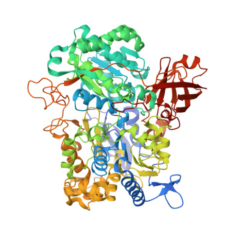
    The Escherichia coli NapA (periplasmic nitrate reductase) contains a [4Fe-4S] cluster and a Mo-bis-molybdopterin guanine dinucleotide cofactor. The NapA holoenzyme associates with a di-heme c-type cytochrome redox partner (NapB). These proteins have been purified and studied by spectropotentiometry, and the structure of NapA has been determined. In contrast to the well characterized heterodimeric NapAB systems ofalpha-proteobacteria, such as Rhodobacter sphaeroides and Paracoccus pantotrophus, the gamma-proteobacterial E. coli NapA and NapB proteins purify independently and not as a tight heterodimeric complex. This relatively weak interaction is reflected in dissociation constants of 15 and 32 mum determined for oxidized and reduced NapAB complexes, respectively. The surface electrostatic potential of E. coli NapA in the apparent NapB binding region is markedly less polar and anionic than that of the alpha-proteobacterial NapA, which may underlie the weaker binding of NapB. The molybdenum ion coordination sphere of E. coli NapA includes two molybdopterin guanine dinucleotide dithiolenes, a protein-derived cysteinyl ligand and an oxygen atom. The Mo-O bond length is 2.6 A, which is indicative of a water ligand. The potential range over which the Mo(6+) state is reduced to the Mo(5+) state in either NapA (between +100 and -100 mV) or the NapAB complex (-150 to -350 mV) is much lower than that reported for R. sphaeroides NapA (midpoint potential Mo(6+/5+) > +350 mV), and the form of the Mo(5+) EPR signal is quite distinct. In E. coli NapA or NapAB, the Mo(5+) state could not be further reduced to Mo(4+). We then propose a catalytic cycle for E. coli NapA in which nitrate binds to the Mo(5+) ion and where a stable des-oxo Mo(6+) species may participate.


  • Organizational Affiliation

    Centre for Metalloprotein Spectroscopy and Biology, University of East Anglia, Norwich NR4 7TJ, United Kingdom.


Macromolecules
Find similar proteins by:  (by identity cutoff)  |  3D Structure
Entity ID: 1
MoleculeChains Sequence LengthOrganismDetailsImage
Periplasmic nitrate reductaseA,
B [auth F]
792Escherichia coli K-12Mutation(s): 0 
Gene Names: napA
EC: 1.7.99.4
UniProt
Find proteins for P33937 (Escherichia coli (strain K12))
Explore P33937 
Go to UniProtKB:  P33937
Entity Groups  
Sequence Clusters30% Identity50% Identity70% Identity90% Identity95% Identity100% Identity
UniProt GroupP33937
Sequence Annotations
Expand
  • Reference Sequence
Small Molecules
Ligands 3 Unique
IDChains Name / Formula / InChI Key2D Diagram3D Interactions
MGD
Query on MGD

Download Ideal Coordinates CCD File 
E [auth A],
F [auth A],
I [auth F],
J [auth F]
2-AMINO-5,6-DIMERCAPTO-7-METHYL-3,7,8A,9-TETRAHYDRO-8-OXA-1,3,9,10-TETRAAZA-ANTHRACEN-4-ONE GUANOSINE DINUCLEOTIDE
C20 H26 N10 O13 P2 S2
VQAGYJCYOLHZDH-ILXWUORBSA-N
SF4
Query on SF4

Download Ideal Coordinates CCD File 
C [auth A],
G [auth F]
IRON/SULFUR CLUSTER
Fe4 S4
LJBDFODJNLIPKO-UHFFFAOYSA-N
6MO
Query on 6MO

Download Ideal Coordinates CCD File 
D [auth A],
H [auth F]
MOLYBDENUM(VI) ION
Mo
HCNGUXXTNNIKCQ-UHFFFAOYSA-N
Experimental Data & Validation

Experimental Data

  • Method: X-RAY DIFFRACTION
  • Resolution: 2.50 Å
  • R-Value Free: 0.243 
  • R-Value Work: 0.181 
  • R-Value Observed: 0.184 
  • Space Group: P 1 21 1
Unit Cell:
Length ( Å )Angle ( ˚ )
a = 69.449α = 90
b = 94.596β = 96.34
c = 131.211γ = 90
Software Package:
Software NamePurpose
SCALAdata scaling
REFMACrefinement
PDB_EXTRACTdata extraction
ADSCdata collection
MOSFLMdata reduction
CCP4data scaling
MOLREPphasing

Structure Validation

View Full Validation Report


Ligand Structure Quality Assessment 


Entry History 

Deposition Data

Revision History  (Full details and data files)

  • Version 1.0: 2006-12-05
    Type: Initial release
  • Version 1.1: 2008-05-01
    Changes: Version format compliance
  • Version 1.2: 2011-07-13
    Changes: Version format compliance
  • Version 1.3: 2023-08-30
    Changes: Data collection, Database references, Derived calculations, Refinement description