2M6N

3D solution structure of EMI1 (Early Mitotic Inhibitor 1)


Experimental Data Snapshot

  • Method: SOLUTION NMR
  • Conformers Calculated: 100 
  • Conformers Submitted: 20 
  • Selection Criteria: target function 

wwPDB Validation   3D Report Full Report


This is version 1.2 of the entry. See complete history


Literature

Electron microscopy structure of human APC/C(CDH1)-EMI1 reveals multimodal mechanism of E3 ligase shutdown.

Frye, J.J.Brown, N.G.Petzold, G.Watson, E.R.Grace, C.R.Nourse, A.Jarvis, M.A.Kriwacki, R.W.Peters, J.M.Stark, H.Schulman, B.A.

(2013) Nat Struct Mol Biol 20: 827-835

  • DOI: https://doi.org/10.1038/nsmb.2593
  • Primary Citation of Related Structures:  
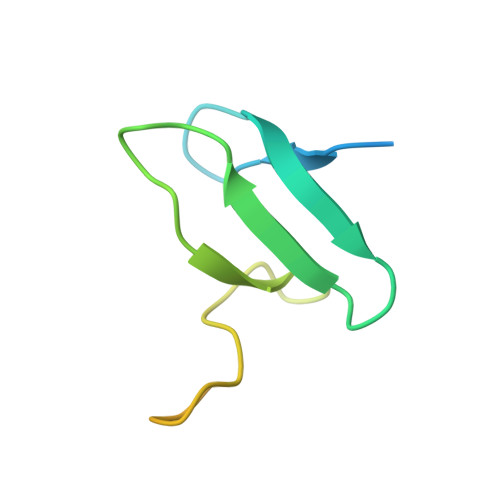
    2M6N

  • PubMed Abstract: 

    The anaphase-promoting complex/cyclosome (APC/C) is a ~1.5-MDa multiprotein E3 ligase enzyme that regulates cell division by promoting timely ubiquitin-mediated proteolysis of key cell-cycle regulatory proteins. Inhibition of human APC/C(CDH1) during interphase by early mitotic inhibitor 1 (EMI1) is essential for accurate coordination of DNA synthesis and mitosis. Here, we report a hybrid structural approach involving NMR, electron microscopy and enzymology, which reveal that EMI1's 143-residue C-terminal domain inhibits multiple APC/C(CDH1) functions. The intrinsically disordered D-box, linker and tail elements, together with a structured zinc-binding domain, bind distinct regions of APC/C(CDH1) to synergistically both block the substrate-binding site and inhibit ubiquitin-chain elongation. The functional importance of intrinsic structural disorder is explained by enabling a small inhibitory domain to bind multiple sites to shut down various functions of a 'molecular machine' nearly 100 times its size.


  • Organizational Affiliation

    1Department of Structural Biology, St. Jude Children's Research Hospital, Memphis, Tennessee, USA.


Macromolecules
Find similar proteins by:  (by identity cutoff)  |  3D Structure
Entity ID: 1
MoleculeChains Sequence LengthOrganismDetailsImage
F-box only protein 586Homo sapiensMutation(s): 0 
Gene Names: FBXO5EMI1FBX5
UniProt & NIH Common Fund Data Resources
Find proteins for Q9UKT4 (Homo sapiens)
Explore Q9UKT4 
Go to UniProtKB:  Q9UKT4
PHAROS:  Q9UKT4
GTEx:  ENSG00000112029 
Entity Groups  
Sequence Clusters30% Identity50% Identity70% Identity90% Identity95% Identity100% Identity
UniProt GroupQ9UKT4
Sequence Annotations
Expand
  • Reference Sequence
Small Molecules
Ligands 1 Unique
IDChains Name / Formula / InChI Key2D Diagram3D Interactions
ZN
Query on ZN

Download Ideal Coordinates CCD File 
B [auth A],
C [auth A]
ZINC ION
Zn
PTFCDOFLOPIGGS-UHFFFAOYSA-N
Experimental Data & Validation

Experimental Data

  • Method: SOLUTION NMR
  • Conformers Calculated: 100 
  • Conformers Submitted: 20 
  • Selection Criteria: target function 

Structure Validation

View Full Validation Report


Entry History 

Deposition Data

Revision History  (Full details and data files)

  • Version 1.0: 2013-05-29
    Type: Initial release
  • Version 1.1: 2013-06-12
    Changes: Database references
  • Version 1.2: 2013-07-17
    Changes: Database references