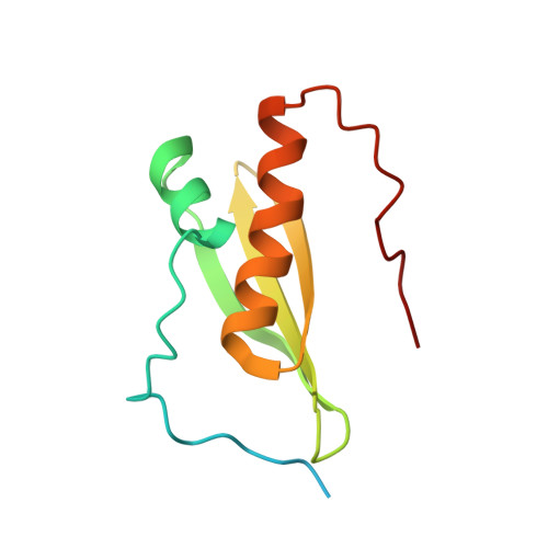
Solution structure of the N-terminal dsRBD of Drosophila ADAR and interaction studies with RNA.
Barraud, P., Heale, B.S., O'Connell, M.A., Allain, F.H.(2012) Biochimie 94: 1499-1509
- PubMed: 22210494
- DOI: https://doi.org/10.1016/j.biochi.2011.12.017
- Primary Citation of Related Structures:
2LJH - PubMed Abstract:
Adenosine deaminases that act on RNA (ADAR) catalyze adenosine to inosine (A-to-I) editing in double-stranded RNA (dsRNA) substrates. Inosine is read as guanosine by the translation machinery; therefore A-to-I editing events in coding sequences may result in recoding genetic information. Whereas vertebrates have two catalytically active enzymes, namely ADAR1 and ADAR2, Drosophila has a single ADAR protein (dADAR) related to ADAR2. The structural determinants controlling substrate recognition and editing of a specific adenosine within dsRNA substrates are only partially understood. Here, we report the solution structure of the N-terminal dsRNA binding domain (dsRBD) of dADAR and use NMR chemical shift perturbations to identify the protein surface involved in RNA binding. Additionally, we show that Drosophila ADAR edits the R/G site in the mammalian GluR-2 pre-mRNA which is naturally modified by both ADAR1 and ADAR2. We then constructed a model showing how dADAR dsRBD1 binds to the GluR-2 R/G stem-loop. This model revealed that most side chains interacting with the RNA sugar-phosphate backbone need only small displacement to adapt for dsRNA binding and are thus ready to bind to their dsRNA target. It also predicts that dADAR dsRBD1 would bind to dsRNA with less sequence specificity than dsRBDs of ADAR2. Altogether, this study gives new insights into dsRNA substrate recognition by Drosophila ADAR.
Organizational Affiliation:
Institute of Molecular Biology and Biophysics, ETH Zurich, Schafmattstrasse 20, CH-8093 Zürich, Switzerland.