NMR analysis of the {alpha}IIb{beta}3 cytoplasmic interaction suggests a mechanism for integrin regulation.
Metcalf, D.G., Moore, D.T., Wu, Y., Kielec, J.M., Molnar, K., Valentine, K.G., Wand, A.J., Bennett, J.S., Degrado, W.F.(2010) Proc Natl Acad Sci U S A 107: 22481-22486
- PubMed: 21156831
- DOI: https://doi.org/10.1073/pnas.1015545107
- Primary Citation of Related Structures:
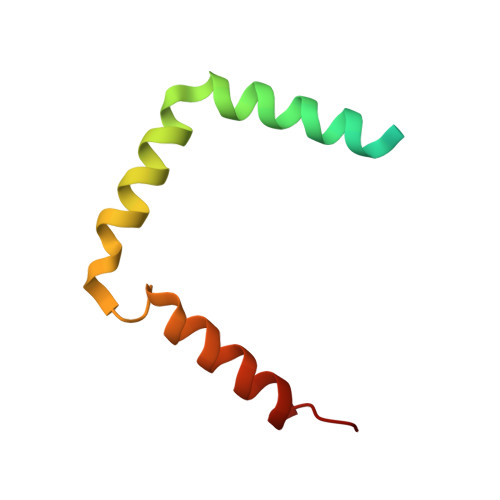
2KV9 - PubMed Abstract:
The integrin αIIbβ3 is a transmembrane (TM) heterodimeric adhesion receptor that exists in equilibrium between resting and active ligand binding conformations. In resting αIIbβ3, the TM and cytoplasmic domains of αIIb and β3 form a heterodimer that constrains αIIbβ3 in its resting conformation. To study the structure and dynamics of the cytoplasmic domain heterodimer, we prepared a disulfide-stabilized complex consisting of portions of the TM domains and the full cytoplasmic domains. NMR and hydrogen-deuterium exchange of this complex in micelles showed that the αIIb cytoplasmic domain is largely disordered, but it interacts with and influences the conformation of the β3 cytoplasmic domain. The β3 cytoplasmic domain consists of a stable proximal helix contiguous with the TM helix and two distal amphiphilic helices. To confirm the NMR structure in a membrane-like environment, we studied the β3 cytoplasmic domain tethered to phospholipid bilayers. Hydrogen-deuterium exchange mass spectrometry, as well as circular dichroism spectroscopy, demonstrated that the β3 cytoplasmic domain becomes more ordered and helical under these conditions, consistent with our NMR results. Further, these experiments suggest that the two distal helices associate with lipid bilayers but undergo fluctuations that would allow rapid binding of cytoplasmic proteins regulating integrin activation, such as talin and kindlin-3. Thus, these results provide a framework for understanding the kinetics and thermodynamics of protein interactions involving integrin cytoplasmic domains and suggest that such interactions act in a concerted fashion to influence integrin stalk separation and exposure of extracellular ligand binding sites.
Organizational Affiliation:
Department of Biochemistry and Biophysics, University of Pennsylvania, Philadelphia, PA 19104, USA.