Structure, Mechanism, and Conformational Dynamics of O-Acetylserine Sulfhydrylase from Salmonella Typhimurium: Comparison of a and B Isozymes.
Chattopadhyay, A., Meier, M., Ivaninskii, S., Burkhard, P., Speroni, F., Campanini, B., Bettati, S., Mozzarelli, A., Rabeh, W.M., Li, L., Cook, P.F.(2007) Biochemistry 46: 8315
- PubMed: 17583914
- DOI: https://doi.org/10.1021/bi602603c
- Primary Citation of Related Structures:
2JC3 - PubMed Abstract:
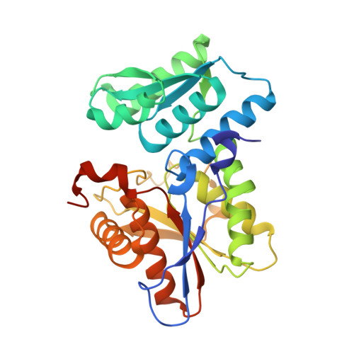
O-Acetylserine sulfhydrylase is a pyridoxal 5'-phosphate-dependent enzyme that catalyzes the final step in the cysteine biosynthetic pathway in enteric bacteria and plants, the replacement of the beta-acetoxy group of O-acetyl-l-serine by a thiol to give l-cysteine. Two isozymes are found in Salmonella typhimurium, with the A-isozyme expressed under aerobic and the B-isozyme expressed under anaerobic conditions. The structure of O-acetylserine sulfhydrylase B has been solved to 2.3 A and exhibits overall a fold very similar to that of the A-isozyme. The main difference between the two isozymes is the more hydrophilic active site of the B-isozyme with two ionizable residues, C280 and D281, replacing the neutral residues S300 and P299, respectively, in the A-isozyme. D281 is above the re face of the cofactor and is within hydrogen-bonding distance to Y286, while C280 is located about 3.4 A from the pyridine nitrogen (N1) of the internal Schiff base. The B-isozyme has a turnover number (V/Et) 12.5-fold higher than the A-isozyme and an approximately 10-fold lower Km for O-acetyl-l-serine. Studies of the first half-reaction by rapid-scanning stopped-flow indicate a first-order conversion of the internal Schiff base to the alpha-aminoacrylate intermediate at any concentration of O-acetyl-l-serine. The Kd values for formation of the external Schiff base with cysteine and serine, obtained by spectral titration, are pH dependent and exhibit a pKa of 7.0-7.5 (for a group that must be unprotonated for optimum binding) with values, above pH 8.0, of about 3.0 and 30.0 mM, respectively. In both cases the neutral enolimine is favored at high pH. Failure to observe the pKa for the alpha-amines of cysteine and serine in the pKESB vs pH profile suggests a compensatory effect resulting from titration of a group on the enzyme with a pKa in the vicinity of the alpha-amine's pKa. The pH dependence of the first-order rate constant for decay of the alpha-aminoacrylate intermediate to give pyruvate and ammonia gives a pKa of about 9 for the active site lysine (K41), a pH unit higher than that of the A-isozyme. The difference in pH dependence of the pKESB for cysteine and serine, the higher pKa for K41, and the preference for the neutral species at high pH compared to the A-isozyme can be explained by titration of C280 to give the thiolate. Subtle conformational differences between O-acetylserine sulfhydrylase A and O-acetylserine sulfhydrylase B are detected by comparing the absorption and emission spectra of the internal aldimine in the absence and presence of the product acetate and of the external aldimine with l-serine. The two isozymes show a different equilibrium distribution of the enolimine and ketoenamine tautomers, likely as a result of a more polar active site for O-acetylserine sulfhydrylase B. The distribution of cofactor tautomers is dramatically affected by the ligation state of the enzyme. In the presence of acetate, which occupies the alpha-carboxylate subsite, the equilibrium between tautomers is shifted toward the ketoenamine tautomer, as a result of a conformational change affecting the structure of the active site. This finding, in agreement with structural data, suggests for the O-acetylserine sulfhydrylase B-isozyme a higher degree of conformational flexibility linked to catalysis.
Organizational Affiliation:
Institute of Materials Science, University of Connecticut, 97 North Eagleville Road, Storrs, Connecticut 06269-3136, USA.