2J9F

Human branched-chain alpha-ketoacid dehydrogenase-decarboxylase E1b


Experimental Data Snapshot

  • Method: X-RAY DIFFRACTION
  • Resolution: 1.88 Å
  • R-Value Free: 0.200 
  • R-Value Work: 0.170 
  • R-Value Observed: 0.171 

wwPDB Validation   3D Report Full Report


Ligand Structure Quality Assessment 


This is version 1.6 of the entry. See complete history


Literature

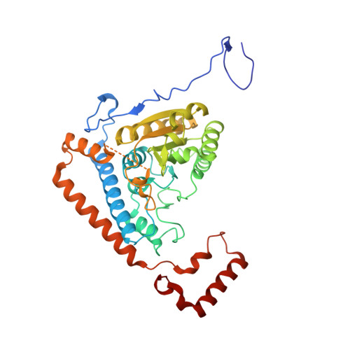
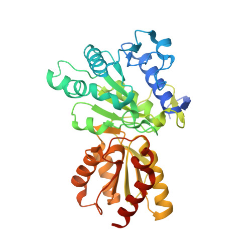
The Two Active Sites in Human Branched-Chain Alpha- Keto Acid Dehydrogenase Operate Independently without an Obligatory Alternating-Site Mechanism.

Li, J.Machius, M.Chuang, J.L.Wynn, R.M.Chuang, D.T.

(2007) J Biol Chem 282: 11904

  • DOI: https://doi.org/10.1074/jbc.M610843200
  • Primary Citation of Related Structures:  
    2J9F

  • PubMed Abstract: 

    A long standing controversy is whether an alternating activesite mechanism occurs during catalysis in thiamine diphosphate (ThDP)-dependent enzymes. We address this question by investigating the ThDP-dependent decarboxylase/dehydrogenase (E1b) component of the mitochondrial branched-chain alpha-keto acid dehydrogenase complex (BCKDC). Our crystal structure reveals that conformations of the two active sites in the human E1b heterotetramer harboring the reaction intermediate are identical. Acidic residues in the core of the E1b heterotetramer, which align with the proton-wire residues proposed to participate in active-site communication in the related pyruvate dehydrogenase from Bacillus stearothermophilus, are mutated. Enzyme kinetic data show that, except in a few cases because of protein misfolding, these alterations are largely without effect on overall activity of BCKDC, ruling out the requirement of a proton-relay mechanism in E1b. BCKDC overall activity is nullified at 50% phosphorylation of E1b, but it is restored to nearly half of the pre-phosphorylation level after dissociation and reconstitution of BCKDC with the same phosphorylated E1b. The results suggest that the abolition of overall activity likely results from the specific geometry of the half-phosphorylated E1b in the BCKDC assembly and not due to a disruption of the alternating active-site mechanism. Finally, we show that a mutant E1b containing only one functional active site exhibits half of the wild-type BCKDC activity, which directly argues against the obligatory communication between active sites. The above results provide evidence that the two active sites in the E1b heterotetramer operate independently during the ThDP-dependent decarboxylation reaction.


  • Organizational Affiliation

    Department of Biochemistry, University of Texas Southwestern Medical Center, Dallas, Texas 75390, USA.


Macromolecules
Find similar proteins by:  (by identity cutoff)  |  3D Structure
Entity ID: 1
MoleculeChains Sequence LengthOrganismDetailsImage
2-OXOISOVALERATE DEHYDROGENASE ALPHA SUBUNIT
A, C
400Homo sapiensMutation(s): 1 
EC: 1.2.4.4
UniProt & NIH Common Fund Data Resources
Find proteins for P12694 (Homo sapiens)
Explore P12694 
Go to UniProtKB:  P12694
PHAROS:  P12694
GTEx:  ENSG00000248098 
Entity Groups  
Sequence Clusters30% Identity50% Identity70% Identity90% Identity95% Identity100% Identity
UniProt GroupP12694
Sequence Annotations
Expand
  • Reference Sequence
Find similar proteins by:  (by identity cutoff)  |  3D Structure
Entity ID: 2
MoleculeChains Sequence LengthOrganismDetailsImage
2-OXOISOVALERATE DEHYDROGENASE BETA SUBUNIT
B, D
350Homo sapiensMutation(s): 0 
EC: 1.2.4.4
UniProt & NIH Common Fund Data Resources
Find proteins for P21953 (Homo sapiens)
Explore P21953 
Go to UniProtKB:  P21953
PHAROS:  P21953
GTEx:  ENSG00000083123 
Entity Groups  
Sequence Clusters30% Identity50% Identity70% Identity90% Identity95% Identity100% Identity
UniProt GroupP21953
Sequence Annotations
Expand
  • Reference Sequence
Small Molecules
Ligands 4 Unique
IDChains Name / Formula / InChI Key2D Diagram3D Interactions
THV
Query on THV

Download Ideal Coordinates CCD File 
G [auth A],
L [auth C]
C2-1-HYDROXY-3-METHYL-PROPYL-THIAMIN DIPHOSPHATE
C16 H26 N4 O8 P2 S
VBABUKBBNKNHAI-UHFFFAOYSA-N
GOL
Query on GOL

Download Ideal Coordinates CCD File 
I [auth B],
N [auth D],
O [auth D]
GLYCEROL
C3 H8 O3
PEDCQBHIVMGVHV-UHFFFAOYSA-N
MN
Query on MN

Download Ideal Coordinates CCD File 
E [auth A],
J [auth C]
MANGANESE (II) ION
Mn
WAEMQWOKJMHJLA-UHFFFAOYSA-N
K
Query on K

Download Ideal Coordinates CCD File 
F [auth A],
H [auth B],
K [auth C],
M [auth D]
POTASSIUM ION
K
NPYPAHLBTDXSSS-UHFFFAOYSA-N
Experimental Data & Validation

Experimental Data

  • Method: X-RAY DIFFRACTION
  • Resolution: 1.88 Å
  • R-Value Free: 0.200 
  • R-Value Work: 0.170 
  • R-Value Observed: 0.171 
  • Space Group: P 32 2 1
Unit Cell:
Length ( Å )Angle ( ˚ )
a = 145.342α = 90
b = 145.342β = 90
c = 138.05γ = 120
Software Package:
Software NamePurpose
REFMACrefinement
HKL-2000data reduction
HKL-2000data scaling
PHASERphasing

Structure Validation

View Full Validation Report


Ligand Structure Quality Assessment 


Entry History 

Deposition Data

Revision History  (Full details and data files)

  • Version 1.0: 2007-02-27
    Type: Initial release
  • Version 1.1: 2011-06-02
    Changes: Version format compliance
  • Version 1.2: 2011-07-13
    Changes: Version format compliance
  • Version 1.3: 2018-10-24
    Changes: Data collection, Source and taxonomy
  • Version 1.4: 2019-05-15
    Changes: Data collection, Experimental preparation
  • Version 1.5: 2019-09-25
    Changes: Data collection, Experimental preparation, Other
  • Version 1.6: 2023-12-13
    Changes: Data collection, Database references, Derived calculations, Refinement description