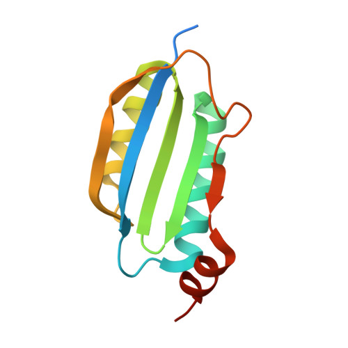
Crystal structures of the AppA BLUF domain photoreceptor provide insights into blue light-mediated signal transduction.
Jung, A., Reinstein, J., Domratcheva, T., Shoeman, R.L., Schlichting, I.(2006) J Mol Biol 362: 717-732
- PubMed: 16949615
- DOI: https://doi.org/10.1016/j.jmb.2006.07.024
- Primary Citation of Related Structures:
2IYG, 2IYI - PubMed Abstract:
Proteins containing a sensor of blue light using FAD (BLUF) domain control diverse cellular processes, such as gene expression, nucleotide metabolism and motility, by relaying blue light signals to distinct output units. Despite its crucial and widespread functions, the mechanism of BLUF signal transduction has remained elusive. We determined crystal structures of the dark-adapted state and of a photo-excited, red-shifted photocycle intermediate of the BLUF unit of AppA, a purple bacterial photoreceptor involved in the light-dependent regulation of photosynthesis gene expression. In contrast to a recently published crystal structure of the AppA BLUF domain determined in the presence of detergent molecules, our structural model of the dark state corresponds well to those reported for the BLUF domains of Tll0078 and BlrB. This establishes that a highly conserved methionine (Met106 in AppA) is next to the active site glutamine (Gln63 in AppA), which is of relevance for the latter's orientation in the dark state and for the mechanism of the photoreaction. The comparison of the dark-adapted and photointermediate state structures shows light-induced conformational alterations, which suggest a path for signal propagation. In particular, we observe a significant movement of the Met106 side-chain. Met106 thereby changes its mode of interaction with Gln63, which supports a light-dependent rotation of the latter. In view of other BLUF structures available, our data further suggest that the hydrogen bond between Asn45 and the backbone carbonyl of His105 breaks upon illumination. The ensuing extensive structural rearrangement of beta-strand 5 is predicted to involve a flip of Met106 out of the flavin-binding pocket and Trp104 moving in to fill the void. We propose that the blue light signal is transmitted towards the surface of the BLUF domain via His44, which serves as a reporter of active site changes.
Organizational Affiliation:
Max-Planck-Institute for Medical Research, Department of Biomolecular Mechanisms, Jahnstr. 29, 69120 Heidelberg, Germany.