Inhibition of metalloprotease botulinum serotype A from a pseudo-peptide binding mode to a small molecule that is active in primary neurons.
Burnett, J.C., Ruthel, G., Stegmann, C.M., Panchal, R.G., Nguyen, T.L., Hermone, A.R., Stafford, R.G., Lane, D.J., Kenny, T.A., McGrath, C.F., Wipf, P., Stahl, A.M., Schmidt, J.J., Gussio, R., Brunger, A.T., Bavari, S.(2007) J Biol Chem 282: 5004-5014
- PubMed: 17092934
- DOI: https://doi.org/10.1074/jbc.M608166200
- Primary Citation of Related Structures:
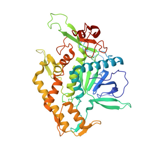
2ISE, 2ISG, 2ISH - PubMed Abstract:
An efficient research strategy integrating empirically guided, structure-based modeling and chemoinformatics was used to discover potent small molecule inhibitors of the botulinum neurotoxin serotype A light chain. First, a modeled binding mode for inhibitor 2-mercapto-3-phenylpropionyl-RATKML (K(i) = 330 nM) was generated, and required the use of a molecular dynamic conformer of the enzyme displaying the reorientation of surface loops bordering the substrate binding cleft. These flexible loops are conformationally variable in x-ray crystal structures, and the model predicted that they were pivotal for providing complementary binding surfaces and solvent shielding for the pseudo-peptide. The docked conformation of 2-mercapto-3-phenylpropionyl-RATKML was then used to refine our pharmacophore for botulinum serotype A light chain inhibition. Data base search queries derived from the pharmacophore were employed to mine small molecule (non-peptidic) inhibitors from the National Cancer Institute's Open Repository. Four of the inhibitors possess K(i) values ranging from 3.0 to 10.0 microM. Of these, NSC 240898 is a promising lead for therapeutic development, as it readily enters neurons, exhibits no neuronal toxicity, and elicits dose-dependent protection of synaptosomal-associated protein (of 25 kDa) in a primary culture of embryonic chicken neurons. Isothermal titration calorimetry showed that the interaction between NSC 240898 and the botulinum A light chain is largely entropy-driven, and occurs with a 1:1 stoichiometry and a dissociation constant of 4.6 microM.
Organizational Affiliation:
Target Structure-based Drug Discovery Group, SAIC-Frederick, Inc., and the National Cancer Institute-Frederick, Frederick, Maryland 21702.