2ILR

Crystal structure of human Fanconi Anemia protein E C-terminal domain


Experimental Data Snapshot

  • Method: X-RAY DIFFRACTION
  • Resolution: 2.00 Å
  • R-Value Free: 0.244 
  • R-Value Work: 0.194 
  • R-Value Observed: 0.196 

wwPDB Validation   3D Report Full Report


This is version 1.3 of the entry. See complete history


Literature

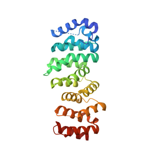
Insights into Fanconi Anaemia from the structure of human FANCE

Nookala, R.K.Hussain, S.Pellegrini, L.

(2007) Nucleic Acids Res 35: 1638-1648

  • DOI: https://doi.org/10.1093/nar/gkm033
  • Primary Citation of Related Structures:  
    2ILR

  • PubMed Abstract: 

    Fanconi Anaemia (FA) is a cancer predisposition disorder characterized by spontaneous chromosome breakage and high cellular sensitivity to genotoxic agents. In response to DNA damage, a multi-subunit assembly of FA proteins, the FA core complex, monoubiquitinates the downstream FANCD2 protein. The FANCE protein plays an essential role in the FA process of DNA repair as the FANCD2-binding component of the FA core complex. Here we report a crystallographic and biological study of human FANCE. The first structure of a FA protein reveals the presence of a repeated helical motif that provides a template for the structural rationalization of other proteins defective in Fanconi Anaemia. The portion of FANCE defined by our crystallographic analysis is sufficient for interaction with FANCD2, yielding structural information into the mode of FANCD2 recruitment to the FA core complex. Disease-associated mutations disrupt the FANCE-FANCD2 interaction, providing structural insight into the molecular mechanisms of FA pathogenesis.


  • Organizational Affiliation

    Department of Biochemistry, University of Cambridge, Tennis Court Road, Cambridge, CB2 1GA, UK.


Macromolecules
Find similar proteins by:  (by identity cutoff)  |  3D Structure
Entity ID: 1
MoleculeChains Sequence LengthOrganismDetailsImage
Fanconi anemia group E protein264Homo sapiensMutation(s): 1 
Gene Names: FANCE
UniProt & NIH Common Fund Data Resources
Find proteins for Q9HB96 (Homo sapiens)
Explore Q9HB96 
Go to UniProtKB:  Q9HB96
PHAROS:  Q9HB96
GTEx:  ENSG00000112039 
Entity Groups  
Sequence Clusters30% Identity50% Identity70% Identity90% Identity95% Identity100% Identity
UniProt GroupQ9HB96
Sequence Annotations
Expand
  • Reference Sequence
Experimental Data & Validation

Experimental Data

  • Method: X-RAY DIFFRACTION
  • Resolution: 2.00 Å
  • R-Value Free: 0.244 
  • R-Value Work: 0.194 
  • R-Value Observed: 0.196 
  • Space Group: P 41 21 2
Unit Cell:
Length ( Å )Angle ( ˚ )
a = 59.291α = 90
b = 59.291β = 90
c = 140.249γ = 90
Software Package:
Software NamePurpose
REFMACrefinement
ADSCdata collection
MOSFLMdata reduction
CCP4data scaling
SHARPphasing

Structure Validation

View Full Validation Report


Entry History 

Deposition Data

Revision History  (Full details and data files)

  • Version 1.0: 2007-02-27
    Type: Initial release
  • Version 1.1: 2008-05-01
    Changes: Version format compliance
  • Version 1.2: 2011-07-13
    Changes: Version format compliance
  • Version 1.3: 2021-11-10
    Changes: Database references