Structure of interleukin 1 alpha at 2.7-A resolution.
Graves, B.J., Hatada, M.H., Hendrickson, W.A., Miller, J.K., Madison, V.S., Satow, Y.(1990) Biochemistry 29: 2679-2684
- PubMed: 2346741
- DOI: https://doi.org/10.1021/bi00463a009
- Primary Citation of Related Structures:
2ILA - PubMed Abstract:
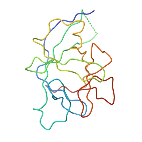
The interleukin 1 (IL-1) family of proteins has a central role in modulating immune and inflammatory responses. Two major IL-1 proteins, designated alpha (IL-1 alpha) and beta (IL-1 beta), are produced by activated macrophages and other cell types. In an effort to understand the similarities and differences in the physicochemical and functional properties of these two proteins, a program was initiated to determine their structures. Crystals of IL-1 alpha were grown, and the three-dimensional structure at 2.7-A resolution was solved. The technique of multiple-wavelength anomalous dispersion (MAD) with the selenomethionine form of IL-1 alpha was utilized in combination with a single mercury derivative to provide the starting phases. Partial refinement of the IL-1 alpha model has been performed as well. The overall structure is composed of 14 beta-strands and a 3(10) helix. The core of this structure is a capped beta-barrell that possesses 3-fold symmetry and displays a topology similar to that observed for IL-1 beta [Priestle, J. P., et al. (1988) EMBO J. 7, 339-343] and soybean trypsin inhibitor (STI) [McLachlan, A. D. (1979) J. Mol. Biol. 133, 557-563]. In this paper, the overall structure of IL-1 alpha and the nature and fidelity of the internal 3-fold symmetry are discussed. Comparisons with IL-1 beta and STI are made within these contexts.
Organizational Affiliation:
Roche Research Center, Hoffmann-La Roche Inc., Nutley, New Jersey 07110.