2IF7

Crystal Structure of NTB-A


Experimental Data Snapshot

  • Method: X-RAY DIFFRACTION
  • Resolution: 3.00 Å
  • R-Value Free: 0.266 
  • R-Value Work: 0.213 
  • R-Value Observed: 0.216 

wwPDB Validation   3D Report Full Report


This is version 1.3 of the entry. See complete history


Literature

NTB-A Receptor Crystal Structure: Insights into Homophilic Interactions in the Signaling Lymphocytic Activation Molecule Receptor Family.

Cao, E.Ramagopal, U.A.Fedorov, A.Fedorov, E.Yan, Q.Lary, J.W.Cole, J.L.Nathenson, S.G.Almo, S.C.

(2006) Immunity 25: 559-570

  • DOI: https://doi.org/10.1016/j.immuni.2006.06.020
  • Primary Citation of Related Structures:  
    2IF7

  • PubMed Abstract: 

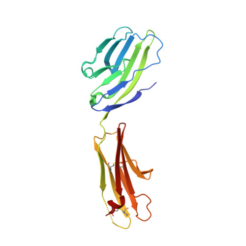
    The signaling lymphocytic activation molecule (SLAM) family includes homophilic and heterophilic receptors that regulate both innate and adaptive immunity. The ectodomains of most SLAM family members are composed of an N-terminal IgV domain and a C-terminal IgC2 domain. NK-T-B-antigen (NTB-A) is a homophilic receptor that stimulates cytotoxicity in natural killer (NK) cells, regulates bactericidal activities in neutrophils, and potentiates T helper 2 (Th2) responses. The 3.0 A crystal structure of the complete NTB-A ectodomain revealed a rod-like monomer that self-associates to form a highly kinked dimer spanning an end-to-end distance of approximately 100 A. The NTB-A homophilic and CD2-CD58 heterophilic dimers show overall structural similarities but differ in detailed organization and physicochemical properties of their respective interfaces. The NTB-A structure suggests a mechanism responsible for binding specificity within the SLAM family and imposes physical constraints relevant to the colocalization of SLAM-family proteins with other signaling molecules in the immunological synapse.


  • Organizational Affiliation

    Department of Cell Biology, Albert Einstein College of Medicine, Bronx, New York 10461, USA.


Macromolecules
Find similar proteins by:  (by identity cutoff)  |  3D Structure
Entity ID: 1
MoleculeChains Sequence LengthOrganismDetailsImage
SLAM family member 6
A, B, C, D
193Homo sapiensMutation(s): 0 
Gene Names: SLAMF6KALI
UniProt & NIH Common Fund Data Resources
Find proteins for Q96DU3 (Homo sapiens)
Explore Q96DU3 
Go to UniProtKB:  Q96DU3
PHAROS:  Q96DU3
GTEx:  ENSG00000162739 
Entity Groups  
Sequence Clusters30% Identity50% Identity70% Identity90% Identity95% Identity100% Identity
UniProt GroupQ96DU3
Sequence Annotations
Expand
  • Reference Sequence
Experimental Data & Validation

Experimental Data

  • Method: X-RAY DIFFRACTION
  • Resolution: 3.00 Å
  • R-Value Free: 0.266 
  • R-Value Work: 0.213 
  • R-Value Observed: 0.216 
  • Space Group: C 1 2 1
Unit Cell:
Length ( Å )Angle ( ˚ )
a = 121.196α = 90
b = 148.452β = 112.86
c = 86.572γ = 90
Software Package:
Software NamePurpose
DENZOdata reduction
SCALEPACKdata scaling
REFMACrefinement
PDB_EXTRACTdata extraction
CBASSdata collection
HKL-2000data reduction
SHELXDphasing
SHELXEmodel building
SOLVEphasing
RESOLVEphasing
Cootmodel building
Omodel building

Structure Validation

View Full Validation Report


Entry History 

Deposition Data

Revision History  (Full details and data files)

  • Version 1.0: 2006-10-17
    Type: Initial release
  • Version 1.1: 2008-05-01
    Changes: Version format compliance
  • Version 1.2: 2011-07-13
    Changes: Advisory, Version format compliance
  • Version 1.3: 2017-10-18
    Changes: Refinement description