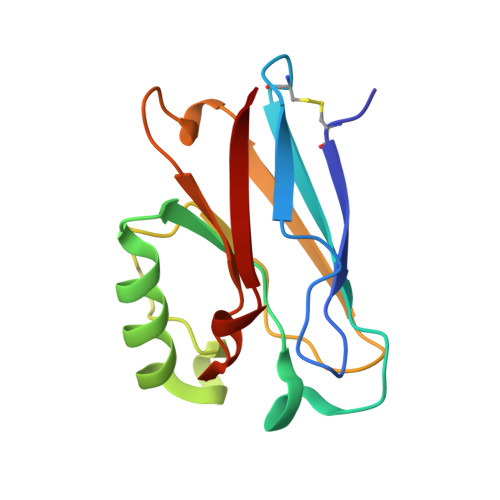
2I7S

Crystal structure of Re(phen)(CO)3 (Thr124His)(His83Gln) Azurin Cu(II) from Pseudomonas aeruginosa


Experimental Data Snapshot

  • Method: X-RAY DIFFRACTION
  • Resolution: 1.35 Å
  • R-Value Free: 0.239 
  • R-Value Work: 0.205 

wwPDB Validation   3D Report Full Report


Ligand Structure Quality Assessment 


This is version 1.4 of the entry. See complete history


Literature

Relaxation dynamics of Pseudomonas aeruginosa Re(I)(CO)3(alpha-diimine)(HisX)+ (X = 83, 107, 109, 124, 126)Cu(II) azurins.

Blanco-Rodriguez, A.M.Busby, M.Ronayne, K.Towrie, M.Gradinaru, C.Sudhamsu, J.Sykora, J.Hof, M.Zalis, S.Di Bilio, A.J.Crane, B.R.Gray, H.B.Vlcek, A.

(2009) J Am Chem Soc 131: 11788-11800

  • DOI: https://doi.org/10.1021/ja902744s
  • Primary Citation of Related Structures:  
    2I7S, 3IBO

  • PubMed Abstract: 

    Photoinduced relaxation processes of five structurally characterized Pseudomonas aeruginosa Re(I)(CO)(3)(alpha-diimine)(HisX) (X = 83, 107, 109, 124, 126)Cu(II) azurins have been investigated by time-resolved (ps-ns) IR spectroscopy and emission spectroscopy. Crystal structures reveal the presence of Re-azurin dimers and trimers that in two cases (X = 107, 124) involve van der Waals interactions between interdigitated diimine aromatic rings. Time-dependent emission anisotropy measurements confirm that the proteins aggregate in mM solutions (D(2)O, KP(i) buffer, pD = 7.1). Excited-state DFT calculations show that extensive charge redistribution in the Re(I)(CO)(3) --> diimine (3)MLCT state occurs: excitation of this (3)MLCT state triggers several relaxation processes in Re-azurins whose kinetics strongly depend on the location of the metallolabel on the protein surface. Relaxation is manifested by dynamic blue shifts of excited-state nu(CO) IR bands that occur with triexponential kinetics: intramolecular vibrational redistribution together with vibrational and solvent relaxation give rise to subps, approximately 2, and 8-20 ps components, while the approximately 10(2) ps kinetics are attributed to displacement (reorientation) of the Re(I)(CO)(3)(phen)(im) unit relative to the peptide chain, which optimizes Coulombic interactions of the Re(I) excited-state electron density with solvated peptide groups. Evidence also suggests that additional segmental movements of Re-bearing beta-strands occur without perturbing the reaction field or interactions with the peptide. Our work demonstrates that time-resolved IR spectroscopy and emission anisotropy of Re(I) carbonyl-diimine complexes are powerful probes of molecular dynamics at or around the surfaces of proteins and protein-protein interfacial regions.


  • Organizational Affiliation

    School of Biological and Chemical Sciences, Queen Mary, University of London, Mile End Road, London E1 4NS, United Kingdom.


Macromolecules
Find similar proteins by:  (by identity cutoff)  |  3D Structure
Entity ID: 1
MoleculeChains Sequence LengthOrganismDetailsImage
Azurin
A, B, C, D
128Pseudomonas aeruginosaMutation(s): 2 
Gene Names: azu
UniProt
Find proteins for P00282 (Pseudomonas aeruginosa (strain ATCC 15692 / DSM 22644 / CIP 104116 / JCM 14847 / LMG 12228 / 1C / PRS 101 / PAO1))
Explore P00282 
Go to UniProtKB:  P00282
Entity Groups  
Sequence Clusters30% Identity50% Identity70% Identity90% Identity95% Identity100% Identity
UniProt GroupP00282
Sequence Annotations
Expand
  • Reference Sequence
Small Molecules
Ligands 3 Unique
IDChains Name / Formula / InChI Key2D Diagram3D Interactions
REP
Query on REP

Download Ideal Coordinates CCD File 
F [auth A],
H [auth B],
K [auth C],
M [auth D]
(1,10 PHENANTHROLINE)-(TRI-CARBON MONOXIDE) RHENIUM (I)
C15 H8 N2 O3 Re
HRZLGVADJDWBIJ-UHFFFAOYSA-N
CON
Query on CON

Download Ideal Coordinates CCD File 
I [auth B]COBALT TETRAAMMINE ION
Co H12 N4
AWWXFUYNCLHXPM-UHFFFAOYSA-N
CU
Query on CU

Download Ideal Coordinates CCD File 
E [auth A],
G [auth B],
J [auth C],
L [auth D]
COPPER (II) ION
Cu
JPVYNHNXODAKFH-UHFFFAOYSA-N
Experimental Data & Validation

Experimental Data

  • Method: X-RAY DIFFRACTION
  • Resolution: 1.35 Å
  • R-Value Free: 0.239 
  • R-Value Work: 0.205 
  • Space Group: P 1
Unit Cell:
Length ( Å )Angle ( ˚ )
a = 33.135α = 82.92
b = 58.98β = 78.11
c = 66.107γ = 76.68
Software Package:
Software NamePurpose
SHELXrefinement
PDB_EXTRACTdata extraction
ADSCdata collection
DENZOdata reduction
SCALEPACKdata scaling
EPMRphasing
SHELXL-97refinement

Structure Validation

View Full Validation Report


Ligand Structure Quality Assessment 


Entry History 

Deposition Data

Revision History  (Full details and data files)

  • Version 1.0: 2006-12-05
    Type: Initial release
  • Version 1.1: 2008-05-01
    Changes: Version format compliance
  • Version 1.2: 2011-07-13
    Changes: Version format compliance
  • Version 1.3: 2021-10-20
    Changes: Database references, Derived calculations
  • Version 1.4: 2023-08-30
    Changes: Data collection, Refinement description