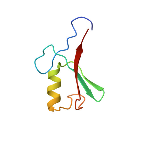
Solution structure of the Hdm2 C2H2C4 RING, a domain critical for ubiquitination of p53.
Kostic, M., Matt, T., Martinez-Yamout, M.A., Dyson, H.J., Wright, P.E.(2006) J Mol Biol 363: 433-450
- PubMed: 16965791
- DOI: https://doi.org/10.1016/j.jmb.2006.08.027
- PubMed Abstract:
Regulation of the transcriptional response to the tumor suppressor p53 occurs at many levels, including control of its transcriptional activity, and of its stability and concentration within the cell. p53 stability is regulated by the protein Hdm2, an E3 ubiquitin ligase that binds to p53 and promotes its ubiquitination and degradation. The C-terminal domain of Hdm2, which is critical for this activity, has been classified as a RING domain on the basis of sequence homology, although it lacks the canonical set of zinc ligands (RING domains typically have C3HC4 or C4C4 zinc coordination). Here, we report the solution structure of the C2H2C4 RING domain of Hdm2(429-491), which reveals a symmetrical dimer with a unique cross-brace zinc-binding scheme. Each subunit has one Cys4 Zn site and one His2Cys2 Zn site. The global fold of each subunit is similar to those reported for other RING domains, with a compact betabetaalphabeta fold, a small hydrophobic core, and two Zn ions, which are essential for maintaining the domain structure. The dimer structure is maintained by an extensive interface that buries a large hydrophobic area on each subunit. It has been proposed that Hdm2 and its homologue HdmX form a stable heterodimer through their RING domains, resulting in a synergistic increase in observed E3 activity. To test this proposal, we prepared an HdmX RING construct and showed by NMR titration that it forms a tight 1:1 complex with the Hdm2 RING. The resonances most perturbed by heterodimer formation are located within the subunit interface of the homodimer, far removed from the surface expected to form the docking site of the E2 ubiquitin-conjugating enzyme, providing a structure-based rationale for the function of the RING domains in p53 ubiquitination.
Organizational Affiliation:
Department of Molecular Biology and Skaggs Institute for Chemical Biology, The Scripps Research Institute, 10550 North Torrey Pines Road, La Jolla, CA 92037, USA.