2H9A

Corrinoid Iron-Sulfur Protein


Experimental Data Snapshot

  • Method: X-RAY DIFFRACTION
  • Resolution: 1.90 Å
  • R-Value Free: 0.260 
  • R-Value Work: 0.203 
  • R-Value Observed: 0.207 

wwPDB Validation   3D Report Full Report


Ligand Structure Quality Assessment 


This is version 1.5 of the entry. See complete history


Literature

Structural insights into methyltransfer reactions of a corrinoid iron-sulfur protein involved in acetyl-CoA synthesis.

Svetlitchnaia, T.Svetlitchnyi, V.Meyer, O.Dobbek, H.

(2006) Proc Natl Acad Sci U S A 103: 14331-14336

  • DOI: https://doi.org/10.1073/pnas.0601420103
  • Primary Citation of Related Structures:  
    2H9A

  • PubMed Abstract: 

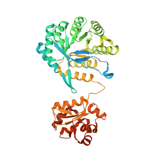
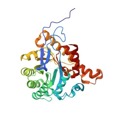
    The cobalt- and iron-containing corrinoid iron-sulfur protein (CoFeSP) is functional in the acetyl-CoA (Ljungdahl-Wood) pathway of autotrophic carbon fixation in various bacteria and archaea, where it is essential for the biosynthesis of acetyl-CoA. CoFeSP acts in two methylation reactions: the transfer of a methyl group from methyltransferase (MeTr)-bound methyltetrahydrofolate to the cob(I)amide of CoFeSP and the transfer of the methyl group of methyl-cob(III)amide to the reduced Ni-Ni-[4Fe-4S] active site cluster A of acetyl-CoA synthase (ACS). We have solved the crystal structure of as-isolated CoFeSP(Ch) from the CO-oxidizing hydrogenogenic bacterium Carboxydothermus hydrogenoformans at 1.9-A resolution. The heterodimeric protein consists of two tightly interacting subunits with pseudo-twofold symmetry. The large CfsA subunit comprises three domains, of which the N-terminal domain binds the [4Fe-4S] cluster, the middle domain is a (betaalpha)(8)-barrel, and the C-terminal domain shows an open fold and binds Cobeta-aqua-(5,6-dimethylbenzimidazolylcobamide) in a "base-off" state without a protein ligand at the cobalt ion. The small CfsB subunit also displays a (betaalpha)(8)-barrel fold and interacts with the upper side of the corrin macrocycle. Structure-based alignments show that both (betaalpha)(8)-barrel domains are related to the MeTr in the acetyl-CoA pathway and to the folate domain of methionine synthase. We suggest that the C-terminal domain of the large subunit is the mobile element that allows the necessary interaction of CoFeSP(Ch) with the active site of ACS(Ch) and the methyltetrahydrofolate carrying MeTr. The conformation in the crystal structure shields the two open coordinations of cobalt and likely represents a resting state.


  • Organizational Affiliation

    Laboratorium Proteinkristallographie, Lehrstuhl für Mikrobiologie, and Bayreuther Zentrum für Molekulare Biowissenschaften, Universität Bayreuth, 95440 Bayreuth, Germany.


Macromolecules
Find similar proteins by:  (by identity cutoff)  |  3D Structure
Entity ID: 1
MoleculeChains Sequence LengthOrganismDetailsImage
Carbon monoxide dehydrogenase corrinoid/iron-sulfur protein, gamma subunit445Carboxydothermus hydrogenoformansMutation(s): 0 
UniProt
Find proteins for Q3ACS3 (Carboxydothermus hydrogenoformans (strain ATCC BAA-161 / DSM 6008 / Z-2901))
Explore Q3ACS3 
Go to UniProtKB:  Q3ACS3
Entity Groups  
Sequence Clusters30% Identity50% Identity70% Identity90% Identity95% Identity100% Identity
UniProt GroupQ3ACS3
Sequence Annotations
Expand
  • Reference Sequence
Find similar proteins by:  (by identity cutoff)  |  3D Structure
Entity ID: 2
MoleculeChains Sequence LengthOrganismDetailsImage
CO dehydrogenase/acetyl-CoA synthase, iron-sulfur protein310Carboxydothermus hydrogenoformansMutation(s): 0 
UniProt
Find proteins for Q3ACS0 (Carboxydothermus hydrogenoformans (strain ATCC BAA-161 / DSM 6008 / Z-2901))
Explore Q3ACS0 
Go to UniProtKB:  Q3ACS0
Entity Groups  
Sequence Clusters30% Identity50% Identity70% Identity90% Identity95% Identity100% Identity
UniProt GroupQ3ACS0
Sequence Annotations
Expand
  • Reference Sequence
Small Molecules
Ligands 3 Unique
IDChains Name / Formula / InChI Key2D Diagram3D Interactions
B12
Query on B12

Download Ideal Coordinates CCD File 
N [auth A]COBALAMIN
C62 H89 Co N13 O14 P
LKVIQTCSMMVGFU-DWSMJLPVSA-N
SF4
Query on SF4

Download Ideal Coordinates CCD File 
O [auth A]IRON/SULFUR CLUSTER
Fe4 S4
LJBDFODJNLIPKO-UHFFFAOYSA-N
IOD
Query on IOD

Download Ideal Coordinates CCD File 
C [auth A]
D [auth A]
E [auth A]
F [auth A]
G [auth A]
C [auth A],
D [auth A],
E [auth A],
F [auth A],
G [auth A],
H [auth A],
I [auth A],
J [auth A],
K [auth A],
L [auth A],
M [auth A],
P [auth B],
Q [auth B],
R [auth B],
S [auth B],
T [auth B]
IODIDE ION
I
XMBWDFGMSWQBCA-UHFFFAOYSA-M
Experimental Data & Validation

Experimental Data

  • Method: X-RAY DIFFRACTION
  • Resolution: 1.90 Å
  • R-Value Free: 0.260 
  • R-Value Work: 0.203 
  • R-Value Observed: 0.207 
  • Space Group: C 1 2 1
Unit Cell:
Length ( Å )Angle ( ˚ )
a = 158.03α = 90
b = 44.27β = 118.63
c = 100.66γ = 90
Software Package:
Software NamePurpose
MAR345data collection
XDSdata reduction
SHARPphasing
CNSrefinement
XDSdata scaling

Structure Validation

View Full Validation Report


Ligand Structure Quality Assessment 


Entry History 

Deposition Data

  • Released Date: 2006-09-26 
  • Deposition Author(s): Dobbek, H.

Revision History  (Full details and data files)

  • Version 1.0: 2006-09-26
    Type: Initial release
  • Version 1.1: 2008-05-01
    Changes: Version format compliance
  • Version 1.2: 2011-07-13
    Changes: Version format compliance
  • Version 1.3: 2012-10-24
    Changes: Non-polymer description
  • Version 1.4: 2017-10-18
    Changes: Refinement description
  • Version 1.5: 2024-02-14
    Changes: Data collection, Database references, Derived calculations