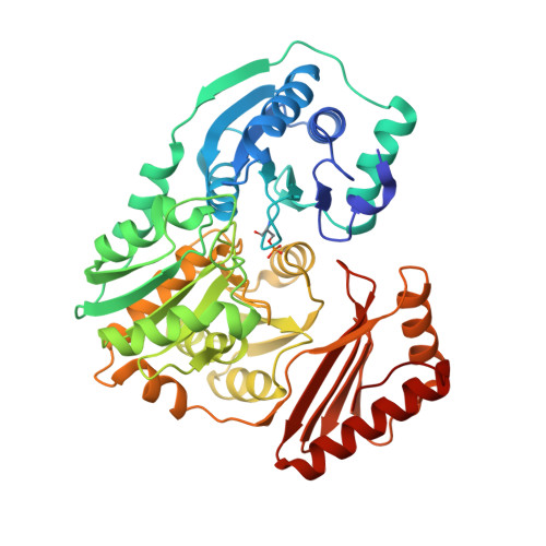
2H4L

Complex of PMM/PGM with ribose 1-phosphate


Experimental Data Snapshot

  • Method: X-RAY DIFFRACTION
  • Resolution: 2.40 Å
  • R-Value Free: 0.259 
  • R-Value Work: 0.186 
  • R-Value Observed: 0.190 

wwPDB Validation   3D Report Full Report


Ligand Structure Quality Assessment 


This is version 2.1 of the entry. See complete history


Literature

Complexes of the enzyme phosphomannomutase/phosphoglucomutase with a slow substrate and an inhibitor.

Regni, C.Shackelford, G.S.Beamer, L.J.

(2006) Acta Crystallogr Sect F Struct Biol Cryst Commun 62: 722-726

  • DOI: https://doi.org/10.1107/S1744309106025887
  • Primary Citation of Related Structures:  
    2H4L, 2H5A

  • PubMed Abstract: 

    Two complexes of the enzyme phosphomannomutase/phosphoglucomutase (PMM/PGM) from Pseudomonas aeruginosa with a slow substrate and with an inhibitor have been characterized by X-ray crystallography. Both ligands induce an interdomain rearrangement in the enzyme that creates a highly buried active site. Comparisons with enzyme-substrate complexes show that the inhibitor xylose 1-phosphate utilizes many of the previously observed enzyme-ligand interactions. In contrast, analysis of the ribose 1-phosphate complex reveals a combination of new and conserved enzyme-ligand interactions for binding. The ability of PMM/PGM to accommodate these two pentose phosphosugars in its active site may be relevant for future efforts towards inhibitor design.


  • Organizational Affiliation

    Department of Structural Biology, St Jude Children's Research Hospital, 332 North Lauderdale M/S 311, Memphis, TN 38105, USA.


Macromolecules
Find similar proteins by:  (by identity cutoff)  |  3D Structure
Entity ID: 1
MoleculeChains Sequence LengthOrganismDetailsImage
Phosphomannomutase/phosphoglucomutaseA [auth X]463Pseudomonas aeruginosaMutation(s): 1 
Gene Names: algC
EC: 5.4.2.8 (PDB Primary Data), 5.4.2.2 (PDB Primary Data)
UniProt
Find proteins for P26276 (Pseudomonas aeruginosa (strain ATCC 15692 / DSM 22644 / CIP 104116 / JCM 14847 / LMG 12228 / 1C / PRS 101 / PAO1))
Explore P26276 
Go to UniProtKB:  P26276
Entity Groups  
Sequence Clusters30% Identity50% Identity70% Identity90% Identity95% Identity100% Identity
UniProt GroupP26276
Sequence Annotations
Expand
  • Reference Sequence
Small Molecules
Ligands 2 Unique
IDChains Name / Formula / InChI Key2D Diagram3D Interactions
R1P
Query on R1P

Download Ideal Coordinates CCD File 
B [auth X]1-O-phosphono-alpha-D-ribofuranose
C5 H11 O8 P
YXJDFQJKERBOBM-TXICZTDVSA-N
ZN
Query on ZN

Download Ideal Coordinates CCD File 
C [auth X]ZINC ION
Zn
PTFCDOFLOPIGGS-UHFFFAOYSA-N
Modified Residues  1 Unique
IDChains TypeFormula2D DiagramParent
SEP
Query on SEP
A [auth X]L-PEPTIDE LINKINGC3 H8 N O6 PSER
Experimental Data & Validation

Experimental Data

  • Method: X-RAY DIFFRACTION
  • Resolution: 2.40 Å
  • R-Value Free: 0.259 
  • R-Value Work: 0.186 
  • R-Value Observed: 0.190 
  • Space Group: P 21 21 21
Unit Cell:
Length ( Å )Angle ( ˚ )
a = 70.717α = 90
b = 74.095β = 90
c = 84.347γ = 90
Software Package:
Software NamePurpose
REFMACrefinement
ADSCdata collection
HKL-2000data scaling

Structure Validation

View Full Validation Report


Ligand Structure Quality Assessment 


Entry History 

Deposition Data

  • Released Date: 2006-08-08 
  • Deposition Author(s): Beamer, L.J.

Revision History  (Full details and data files)

  • Version 1.0: 2006-08-08
    Type: Initial release
  • Version 1.1: 2008-05-01
    Changes: Version format compliance
  • Version 1.2: 2011-07-13
    Changes: Advisory, Version format compliance
  • Version 2.0: 2020-07-29
    Type: Remediation
    Reason: Carbohydrate remediation
    Changes: Atomic model, Data collection, Database references, Derived calculations, Structure summary
  • Version 2.1: 2023-08-30
    Changes: Data collection, Database references, Refinement description, Structure summary