2GX2

Crystal structural and functional analysis of GFP-like fluorescent protein Dronpa


Experimental Data Snapshot

  • Method: X-RAY DIFFRACTION
  • Resolution: 1.80 Å
  • R-Value Free: 0.209 
  • R-Value Work: 0.182 
  • R-Value Observed: 0.182 

wwPDB Validation   3D Report Full Report


This is version 2.0 of the entry. See complete history


Literature

Structural characterization of the photoswitchable fluorescent protein Dronpa-C62S

Nam, K.-H.Kwon, O.Y.Sugiyama, K.Lee, W.-H.Kim, Y.K.Song, H.K.Kim, E.E.Park, S.-Y.Jeon, H.Hwang, K.Y.

(2007) Biochem Biophys Res Commun 354: 962-967

  • DOI: https://doi.org/10.1016/j.bbrc.2007.01.086
  • Primary Citation of Related Structures:  
    2GX0, 2GX2

  • PubMed Abstract: 

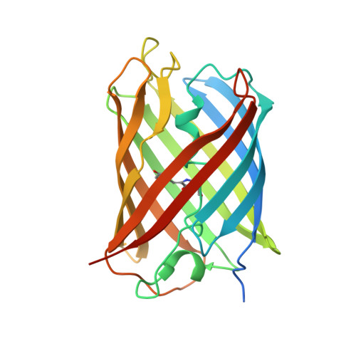
    The photoswitching behavior of green fluorescent proteins (GFPs) or GFP-like proteins is increasingly recognized as a new technique for optical marking. Recently, Ando and his colleagues developed a new green fluorescent protein Dronpa, which possesses the unique photochromic property of being photoswitchable in a non-destructive manner. To better understand this mechanism, we determined the crystal structures of a new GFP Dronpa and its mutant C62S, at 1.9 Angstroms and 1.8 Angstroms, respectively. Determination of the structures demonstrates that a unique hydrogen-bonding network and the sulfur atom of the chromophore are critical to the photoswitching property of Dronpa. Reversible photoswitching was lost in cells expressing the Dronpa-C62S upon repetitive irradiation compared to the native protein. Structural and mutational analyses reveal the chemical basis for the functional properties of photoswitchable fluorescent proteins and provide the basis for subsequent coherent engineering of this subfamily of Dronpa homologs.


  • Organizational Affiliation

    Division of Biotechnology, College of Life Sciences & Biotechnology, Korea University, Anam-Dong, Sungbuk-Gu, Seoul, South Korea.


Macromolecules
Find similar proteins by:  (by identity cutoff)  |  3D Structure
Entity ID: 1
MoleculeChains Sequence LengthOrganismDetailsImage
fluorescent protein Dronpa
A, B, C, D, E
A, B, C, D, E, F, G, H, I, J, K, L
241Echinophyllia sp. SC22Mutation(s): 1 
UniProt
Find proteins for Q5TLG6 (Echinophyllia sp. SC22)
Explore Q5TLG6 
Go to UniProtKB:  Q5TLG6
Entity Groups  
Sequence Clusters30% Identity50% Identity70% Identity90% Identity95% Identity100% Identity
UniProt GroupQ5TLG6
Sequence Annotations
Expand
  • Reference Sequence
Small Molecules
Ligands 1 Unique
IDChains Name / Formula / InChI Key2D Diagram3D Interactions
MG
Query on MG

Download Ideal Coordinates CCD File 
AA [auth C]
AB [auth F]
BA [auth C]
BB [auth G]
CA [auth C]
AA [auth C],
AB [auth F],
BA [auth C],
BB [auth G],
CA [auth C],
CB [auth G],
DA [auth C],
DB [auth G],
EA [auth C],
EB [auth H],
FA [auth C],
FB [auth H],
GA [auth C],
GB [auth H],
HA [auth C],
HB [auth H],
IA [auth D],
IB [auth I],
JA [auth D],
JB [auth I],
KA [auth D],
KB [auth I],
LA [auth D],
LB [auth I],
M [auth A],
MA [auth D],
MB [auth J],
N [auth A],
NA [auth D],
NB [auth K],
O [auth A],
OA [auth D],
OB [auth K],
P [auth A],
PA [auth E],
PB [auth K],
Q [auth A],
QA [auth E],
QB [auth K],
R [auth A],
RA [auth E],
RB [auth K],
S [auth B],
SA [auth E],
SB [auth K],
T [auth B],
TA [auth E],
TB [auth L],
U [auth B],
UA [auth E],
UB [auth L],
V [auth B],
VA [auth E],
VB [auth L],
W [auth B],
WA [auth F],
X [auth B],
XA [auth F],
Y [auth B],
YA [auth F],
Z [auth B],
ZA [auth F]
MAGNESIUM ION
Mg
JLVVSXFLKOJNIY-UHFFFAOYSA-N
Modified Residues  1 Unique
IDChains TypeFormula2D DiagramParent
GYS
Query on GYS
A, B, C, D, E
A, B, C, D, E, F, G, H, I, J, K, L
L-PEPTIDE LINKINGC14 H15 N3 O5SER, TYR, GLY
Experimental Data & Validation

Experimental Data

  • Method: X-RAY DIFFRACTION
  • Resolution: 1.80 Å
  • R-Value Free: 0.209 
  • R-Value Work: 0.182 
  • R-Value Observed: 0.182 
  • Space Group: P 1
Unit Cell:
Length ( Å )Angle ( ˚ )
a = 73.091α = 61.17
b = 106.16β = 70.85
c = 110.07γ = 86.21
Software Package:
Software NamePurpose
CNSrefinement
ADSCdata collection
HKL-2000data scaling
X-PLORphasing

Structure Validation

View Full Validation Report


Entry History 

Deposition Data

Revision History  (Full details and data files)

  • Version 1.0: 2007-05-08
    Type: Initial release
  • Version 1.1: 2008-05-01
    Changes: Version format compliance
  • Version 1.2: 2011-07-13
    Changes: Version format compliance
  • Version 1.3: 2023-10-25
    Changes: Data collection, Database references, Derived calculations, Refinement description
  • Version 2.0: 2023-11-15
    Changes: Atomic model, Data collection, Derived calculations