2GH6

Crystal structure of a HDAC-like protein with 9,9,9-trifluoro-8-oxo-N-phenylnonan amide bound


Experimental Data Snapshot

  • Method: X-RAY DIFFRACTION
  • Resolution: 2.20 Å
  • R-Value Free: 0.214 
  • R-Value Work: 0.158 
  • R-Value Observed: 0.161 

wwPDB Validation   3D Report Full Report


Ligand Structure Quality Assessment 


This is version 1.3 of the entry. See complete history


Literature

Complex structure of a bacterial class 2 histone deacetylase homologue with a trifluoromethylketone inhibitor.

Nielsen, T.K.Hildmann, C.Riester, D.Wegener, D.Schwienhorst, A.Ficner, R.

(2007) Acta Crystallogr Sect F Struct Biol Cryst Commun 63: 270-273

  • DOI: https://doi.org/10.1107/S1744309107012377
  • Primary Citation of Related Structures:  
    2GH6

  • PubMed Abstract: 

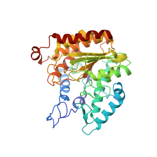
    Histone deacetylases (HDACs) have emerged as attractive targets in anticancer drug development. To date, a number of HDAC inhibitors have been developed and most of them are hydroxamic acid derivatives, typified by suberoylanilide hydroxamic acid (SAHA). Not surprisingly, structural information that can greatly enhance the design of novel HDAC inhibitors is so far only available for hydroxamic acids in complex with HDAC or HDAC-like enzymes. Here, the first structure of an enzyme complex with a nonhydroxamate HDAC inhibitor is presented. The structure of the trifluoromethyl ketone inhibitor 9,9,9-trifluoro-8-oxo-N-phenylnonanamide in complex with bacterial FB188 HDAH (histone deacetylase-like amidohydrolase from Bordetella/Alcaligenes strain FB188) has been determined. HDAH reveals high sequential and functional homology to human class 2 HDACs and a high structural homology to human class 1 HDACs. Comparison with the structure of HDAH in complex with SAHA reveals that the two inhibitors superimpose well. However, significant differences in binding to the active site of HDAH were observed. In the presented structure the O atom of the trifluoromethyl ketone moiety is within binding distance of the Zn atom of the enzyme and the F atoms participate in interactions with the enzyme, thereby involving more amino acids in enzyme-inhibitor binding.


  • Organizational Affiliation

    Abteilung für Molekulare Strukturbiologie, Institut für Mikrobiologie und Genetik and GZMB, Justus-von-Liebig Weg 11, 37077 Göttingen, Germany.


Macromolecules
Find similar proteins by:  (by identity cutoff)  |  3D Structure
Entity ID: 1
MoleculeChains Sequence LengthOrganismDetailsImage
Histone deacetylase-like amidohydrolase
A, B, C, D
369Alcaligenaceae bacterium FB188Mutation(s): 0 
Gene Names: hdaHhdaH1
EC: 3.5.1
UniProt
Find proteins for Q70I53 (Alcaligenes sp. (strain DSM 11172))
Explore Q70I53 
Go to UniProtKB:  Q70I53
Entity Groups  
Sequence Clusters30% Identity50% Identity70% Identity90% Identity95% Identity100% Identity
UniProt GroupQ70I53
Sequence Annotations
Expand
  • Reference Sequence
Small Molecules
Ligands 3 Unique
IDChains Name / Formula / InChI Key2D Diagram3D Interactions
CF3
Query on CF3

Download Ideal Coordinates CCD File 
H [auth A],
L [auth B],
P [auth C],
T [auth D]
9,9,9-TRIFLUORO-8-OXO-N-PHENYLNONANAMIDE
C15 H18 F3 N O2
KRCXZGYVOZSCSF-UHFFFAOYSA-N
ZN
Query on ZN

Download Ideal Coordinates CCD File 
E [auth A],
I [auth B],
M [auth C],
Q [auth D]
ZINC ION
Zn
PTFCDOFLOPIGGS-UHFFFAOYSA-N
K
Query on K

Download Ideal Coordinates CCD File 
F [auth A]
G [auth A]
J [auth B]
K [auth B]
N [auth C]
F [auth A],
G [auth A],
J [auth B],
K [auth B],
N [auth C],
O [auth C],
R [auth D],
S [auth D]
POTASSIUM ION
K
NPYPAHLBTDXSSS-UHFFFAOYSA-N
Binding Affinity Annotations 
IDSourceBinding Affinity
CF3 PDBBind:  2GH6 IC50: 1.12e+4 (nM) from 1 assay(s)
Experimental Data & Validation

Experimental Data

  • Method: X-RAY DIFFRACTION
  • Resolution: 2.20 Å
  • R-Value Free: 0.214 
  • R-Value Work: 0.158 
  • R-Value Observed: 0.161 
  • Space Group: P 1 21 1
Unit Cell:
Length ( Å )Angle ( ˚ )
a = 68.507α = 90
b = 94.897β = 104.28
c = 123.301γ = 90
Software Package:
Software NamePurpose
HKL-2000data reduction
MOLREPphasing
REFMACrefinement
HKL-2000data scaling

Structure Validation

View Full Validation Report


Ligand Structure Quality Assessment 


Entry History 

Deposition Data

Revision History  (Full details and data files)

  • Version 1.0: 2007-03-20
    Type: Initial release
  • Version 1.1: 2008-05-01
    Changes: Version format compliance
  • Version 1.2: 2011-07-13
    Changes: Derived calculations, Version format compliance
  • Version 1.3: 2023-08-30
    Changes: Data collection, Database references, Derived calculations, Refinement description