


2GD4
Crystal Structure of the Antithrombin-S195A Factor Xa-Pentasaccharide Complex
- PDB DOI: https://doi.org/10.2210/pdb2GD4/pdb
- Classification: HYDROLASE
- Organism(s): Homo sapiens
- Expression System: Escherichia coli
- Mutation(s): Yes
- Deposited: 2006-03-15 Released: 2006-05-09
Experimental Data Snapshot
- Method: X-RAY DIFFRACTION
- Resolution: 3.30 Å
- R-Value Free: 0.298
- R-Value Work: 0.247

This is version 2.3 of the entry. See complete history.

Macromolecules
Find similar proteins by:
(by identity cutoff) | 3D Structure
Entity ID: 1 | |||||
|---|---|---|---|---|---|
| Molecule | Chains | Sequence Length | Organism | Details | Image |
| Coagulation factor X, Stuart factor, Stuart-Prower factor, Contains: Factor X light chain; Factor X heavy chain; Activated factor Xa heavy chain | A [auth L], D [auth A] | 58 | Homo sapiens | Mutation(s): 0 Gene Names: F10 EC: 3.4.21.6 | 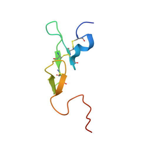 |
UniProt & NIH Common Fund Data Resources | |||||
Find proteins for P00742 (Homo sapiens) Explore P00742 Go to UniProtKB: P00742 | |||||
PHAROS: P00742 GTEx: ENSG00000126218 | |||||
Entity Groups | |||||
| Sequence Clusters | 30% Identity50% Identity70% Identity90% Identity95% Identity100% Identity | ||||
| UniProt Group | P00742 | ||||
Sequence AnnotationsExpand | |||||
| |||||
Find similar proteins by:
(by identity cutoff) | 3D Structure
Entity ID: 2 | |||||
|---|---|---|---|---|---|
| Molecule | Chains | Sequence Length | Organism | Details | Image |
| Coagulation factor, Stuart factor, Stuart-Prower factor, Contains: Factor X light chain; Factor X heavy chain; Activated factor Xa heavy chain | B [auth H], E [auth B] | 241 | Homo sapiens | Mutation(s): 1 Gene Names: F10 EC: 3.4.21.6 |  |
UniProt & NIH Common Fund Data Resources | |||||
Find proteins for P00742 (Homo sapiens) Explore P00742 Go to UniProtKB: P00742 | |||||
PHAROS: P00742 GTEx: ENSG00000126218 | |||||
Entity Groups | |||||
| Sequence Clusters | 30% Identity50% Identity70% Identity90% Identity95% Identity100% Identity | ||||
| UniProt Group | P00742 | ||||
Sequence AnnotationsExpand | |||||
| |||||
Find similar proteins by:
(by identity cutoff) | 3D Structure
Entity ID: 3 | |||||
|---|---|---|---|---|---|
| Molecule | Chains | Sequence Length | Organism | Details | Image |
| Antithrombin-III | C [auth I], F [auth C] | 443 | Homo sapiens | Mutation(s): 4 Gene Names: SERPINC1, AT3 | 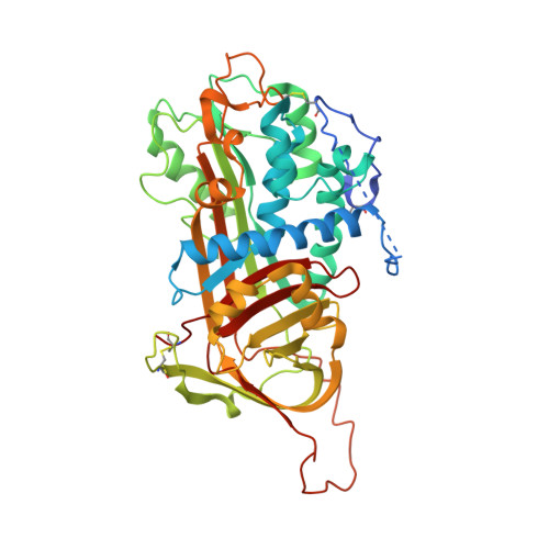 |
UniProt & NIH Common Fund Data Resources | |||||
Find proteins for P01008 (Homo sapiens) Explore P01008 Go to UniProtKB: P01008 | |||||
PHAROS: P01008 GTEx: ENSG00000117601 | |||||
Entity Groups | |||||
| Sequence Clusters | 30% Identity50% Identity70% Identity90% Identity95% Identity100% Identity | ||||
| UniProt Group | P01008 | ||||
Sequence AnnotationsExpand | |||||
| |||||
Oligosaccharides
Entity ID: 4 | |||||
|---|---|---|---|---|---|
| Molecule | Chains | Length | 2D Diagram | Glycosylation | 3D Interactions |
| alpha-D-mannopyranose-(1-2)-alpha-D-mannopyranose-(1-3)-[alpha-D-mannopyranose-(1-6)]beta-D-mannopyranose-(1-4)-2-acetamido-2-deoxy-beta-D-glucopyranose-(1-4)-2-acetamido-2-deoxy-beta-D-glucopyranose | G [auth D] | 6 |  | N-Glycosylation | |
Glycosylation Resources | |||||
GlyTouCan: G56014GC GlyCosmos: G56014GC GlyGen: G56014GC | |||||
Entity ID: 5 | |||||
|---|---|---|---|---|---|
| Molecule | Chains | Length | 2D Diagram | Glycosylation | 3D Interactions |
| 2-acetamido-2-deoxy-beta-D-glucopyranose-(1-4)-2-acetamido-2-deoxy-beta-D-glucopyranose | H [auth E] | 2 |  | N-Glycosylation | |
Glycosylation Resources | |||||
GlyTouCan: G42666HT GlyCosmos: G42666HT GlyGen: G42666HT | |||||
Entity ID: 6 | |||||
|---|---|---|---|---|---|
| Molecule | Chains | Length | 2D Diagram | Glycosylation | 3D Interactions |
| 2-deoxy-6-O-sulfo-2-(sulfoamino)-alpha-D-glucopyranose-(1-4)-beta-D-glucopyranuronic acid-(1-4)-2-deoxy-3,6-di-O-sulfo-2-(sulfoamino)-alpha-D-glucopyranose-(1-4)-2-O-sulfo-alpha-L-idopyranuronic acid-(1-4)-methyl 2-deoxy-6-O-sulfo-2-(sulfoamino)-alpha-D-glucopyranoside | I [auth F], J [auth G] | 5 |  | N/A | |
Glycosylation Resources | |||||
GlyTouCan: G25282IW GlyCosmos: G25282IW | |||||
Small Molecules
| Ligands 2 Unique | |||||
|---|---|---|---|---|---|
| ID | Chains | Name / Formula / InChI Key | 2D Diagram | 3D Interactions | |
| NAG Query on NAG | L [auth I], M [auth I], O [auth C], P [auth C] | 2-acetamido-2-deoxy-beta-D-glucopyranose C8 H15 N O6 OVRNDRQMDRJTHS-FMDGEEDCSA-N |  | ||
| CA Query on CA | K [auth H], N [auth B] | CALCIUM ION Ca BHPQYMZQTOCNFJ-UHFFFAOYSA-N |  | ||
Biologically Interesting Molecules (External Reference) 1 Unique
Entity ID: 6 | |||||
|---|---|---|---|---|---|
| ID | Chains | Name | Type/Class | 2D Diagram | 3D Interactions |
| PRD_900028 Query on PRD_900028 | I [auth F], J [auth G] | fondaparinux | Oligosaccharide / Anticoagulant |  | |
Experimental Data & Validation
Experimental Data
- Method: X-RAY DIFFRACTION
- Resolution: 3.30 Å
- R-Value Free: 0.298
- R-Value Work: 0.247
- Space Group: C 1 2 1
Unit Cell:
| Length ( Å ) | Angle ( ˚ ) |
|---|---|
| a = 220.263 | α = 90 |
| b = 60.588 | β = 113.14 |
| c = 156.174 | γ = 90 |
| Software Name | Purpose |
|---|---|
| SCALA | data scaling |
| PHASER | phasing |
| CNS | refinement |
| PDB_EXTRACT | data extraction |
| ADSC | data collection |
| CCP4 | data scaling |
Entry History
Deposition Data
- Released Date: 2006-05-09 Deposition Author(s): Johnson, D.J., Li, W., Adams, T.E., Huntington, J.A.
Revision History (Full details and data files)
- Version 1.0: 2006-05-09
Type: Initial release - Version 1.1: 2008-05-01
Changes: Version format compliance - Version 1.2: 2011-07-13
Changes: Non-polymer description, Version format compliance - Version 1.3: 2017-07-12
Changes: Advisory, Structure summary - Version 2.0: 2020-07-29
Type: Remediation
Reason: Carbohydrate remediation
Changes: Advisory, Atomic model, Data collection, Database references, Derived calculations, Non-polymer description, Source and taxonomy, Structure summary - Version 2.1: 2021-10-20
Changes: Advisory, Database references, Structure summary - Version 2.2: 2023-08-30
Changes: Data collection, Refinement description - Version 2.3: 2024-03-13
Changes: Source and taxonomy, Structure summary
