2G6E

Structure of cyclized F64L S65A Y66S GFP variant


Experimental Data Snapshot

  • Method: X-RAY DIFFRACTION
  • Resolution: 1.30 Å
  • R-Value Free: 0.187 
  • R-Value Work: 0.131 

wwPDB Validation   3D Report Full Report


This is version 1.5 of the entry. See complete history


Literature

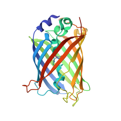
Understanding GFP Posttranslational Chemistry: Structures of Designed Variants that Achieve Backbone Fragmentation, Hydrolysis, and Decarboxylation.

Barondeau, D.P.Kassmann, C.J.Tainer, J.A.Getzoff, E.D.

(2006) J Am Chem Soc 128: 4685-4693

  • DOI: https://doi.org/10.1021/ja056635l
  • Primary Citation of Related Structures:  
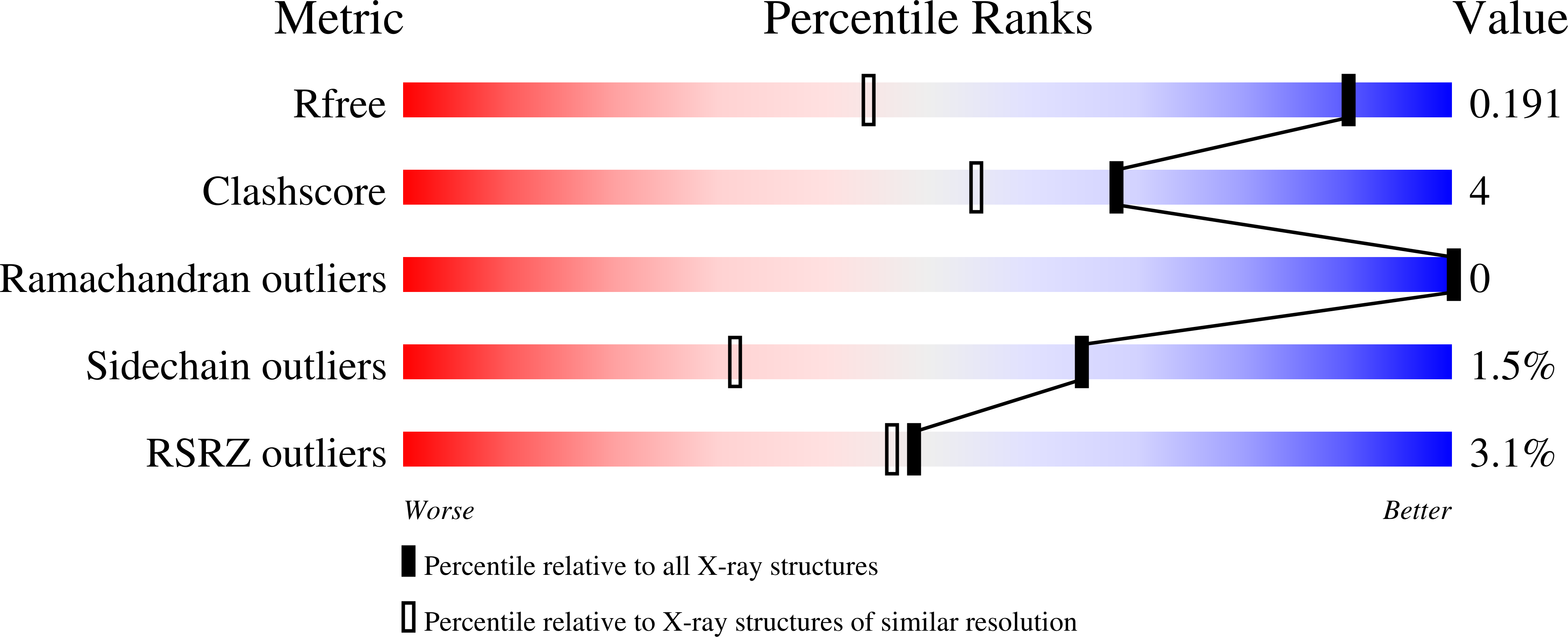
    2G16, 2G2S, 2G3D, 2G5Z, 2G6E

  • PubMed Abstract: 

    The green fluorescent protein (GFP) creates a fluorophore out of three sequential amino acids by promoting spontaneous posttranslational modifications. Here, we use high-resolution crystallography to characterize GFP variants that not only undergo peptide backbone cyclization but additional denaturation-induced peptide backbone fragmentation, native peptide hydrolysis, and decarboxylation reactions. Our analyses indicate that architectural features that favor GFP peptide cyclization also drive peptide hydrolysis. These results are relevant for the maturation pathways of GFP homologues, such as the kindling fluorescent protein and the Kaede protein, which use backbone cleavage to red-shift the spectral properties of their chromophores. We further propose a photochemical mechanism for the decarboxylation reaction, supporting a role for the GFP protein environment in facilitating radical formation and one-electron chemistry, which may be important in activating oxygen for the oxidation step of chromophore biosynthesis. Together, our results characterize GFP posttranslational modification chemistry with implications for the energetic landscape of backbone cyclization and subsequent reactions, and for the rational design of predetermined spontaneous backbone cyclization and cleavage reactions.


  • Organizational Affiliation

    Department of Molecular Biology, The Skaggs Institute for Chemical Biology, The Scripps Research Institute, 10550 North Torrey Pines Road, La Jolla, California 92037, USA.


Macromolecules
Find similar proteins by:  (by identity cutoff)  |  3D Structure
Entity ID: 1
MoleculeChains Sequence LengthOrganismDetailsImage
Green fluorescent protein237Aequorea victoriaMutation(s): 5 
Gene Names: GFP
UniProt
Find proteins for P42212 (Aequorea victoria)
Explore P42212 
Go to UniProtKB:  P42212
Entity Groups  
Sequence Clusters30% Identity50% Identity70% Identity90% Identity95% Identity100% Identity
UniProt GroupP42212
Sequence Annotations
Expand
  • Reference Sequence
Small Molecules
Ligands 1 Unique
IDChains Name / Formula / InChI Key2D Diagram3D Interactions
MG
Query on MG

Download Ideal Coordinates CCD File 
B [auth A]MAGNESIUM ION
Mg
JLVVSXFLKOJNIY-UHFFFAOYSA-N
Modified Residues  1 Unique
IDChains TypeFormula2D DiagramParent
CRX
Query on CRX
A
L-PEPTIDE LINKINGC8 H13 N3 O4ALA, SER, GLY
Experimental Data & Validation

Experimental Data

  • Method: X-RAY DIFFRACTION
  • Resolution: 1.30 Å
  • R-Value Free: 0.187 
  • R-Value Work: 0.131 
  • Space Group: P 21 21 21
Unit Cell:
Length ( Å )Angle ( ˚ )
a = 51.09α = 90
b = 62.62β = 90
c = 71.36γ = 90
Software Package:
Software NamePurpose
DENZOdata reduction
SCALEPACKdata scaling
AMoREphasing
SHELXL-97refinement

Structure Validation

View Full Validation Report


Entry History 

Deposition Data

Revision History  (Full details and data files)

  • Version 1.0: 2006-04-18
    Type: Initial release
  • Version 1.1: 2008-05-01
    Changes: Version format compliance
  • Version 1.2: 2011-07-13
    Changes: Version format compliance
  • Version 1.3: 2021-10-20
    Changes: Database references, Derived calculations
  • Version 1.4: 2023-08-30
    Changes: Data collection, Refinement description
  • Version 1.5: 2023-11-15
    Changes: Data collection, Derived calculations