Structure of the Sulfolobus solfataricus alpha-Glucosidase: Implications for Domain Conservation and Substrate Recognition in GH31.
Ernst, H.A., Lo Leggio, L., Willemoes, M., Leonard, G., Blum, P., Larsen, S.(2006) J Mol Biol 358: 1106-1124
- PubMed: 16580018
- DOI: https://doi.org/10.1016/j.jmb.2006.02.056
- Primary Citation of Related Structures:
2G3M, 2G3N - PubMed Abstract:
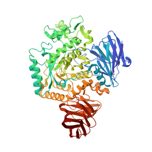
The crystal structure of alpha-glucosidase MalA from Sulfolobus solfataricus has been determined at 2.5Angstrom resolution. It provides a structural model for enzymes representing the major specificity in glycoside hydrolase family 31 (GH31), including alpha-glucosidases from higher organisms, involved in glycogen degradation and glycoprotein processing. The structure of MalA shows clear differences from the only other structure known from GH31, alpha-xylosidase YicI. MalA and YicI share only 23% sequence identity. Although the two enzymes display a similar domain structure and both form hexamers, their structures differ significantly in quaternary organization: MalA is a dimer of trimers, YicI a trimer of dimers. MalA and YicI also differ in their substrate specificities, as shown by kinetic measurements on model chromogenic substrates. In addition, MalA has a clear preference for maltose (Glc-alpha1,4-Glc), whereas YicI prefers isoprimeverose (Xyl-alpha1,6-Glc). The structural origin of this difference occurs in the -1 subsite where MalA residues Asp251 and Trp284 could interact with OH6 of the substrate. The structure of MalA in complex with beta-octyl-glucopyranoside has been determined. It reveals Arg400, Asp87, Trp284, Met321 and Phe327 as invariant residues forming the +1 subsite in the GH31 alpha-glucosidases. Structural comparisons with other GH families suggest that the GH31 enzymes belong to clan GH-D.
Organizational Affiliation:
Biophysical Chemistry Group, Department of Chemistry, University of Copenhagen, Universitetsparken 5, DK-2100 Copenhagen Ø, Denmark.