2G09

X-ray structure of mouse pyrimidine 5'-nucleotidase type 1, product complex


Experimental Data Snapshot

  • Method: X-RAY DIFFRACTION
  • Resolution: 2.10 Å
  • R-Value Free: 0.198 
  • R-Value Work: 0.150 
  • R-Value Observed: 0.153 

wwPDB Validation   3D Report Full Report


Ligand Structure Quality Assessment 


This is version 1.5 of the entry. See complete history


Literature

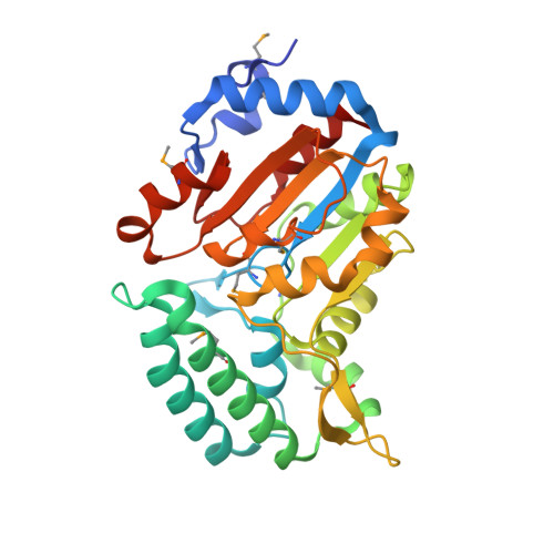
Structure of pyrimidine 5'-nucleotidase type 1. Insight into mechanism of action and inhibition during lead poisoning.

Bitto, E.Bingman, C.A.Wesenberg, G.E.McCoy, J.G.Phillips, G.N.

(2006) J Biol Chem 281: 20521-20529

  • DOI: https://doi.org/10.1074/jbc.M602000200
  • Primary Citation of Related Structures:  
    2BDU, 2G06, 2G07, 2G08, 2G09, 2G0A

  • PubMed Abstract: 

    Eukaryotic pyrimidine 5'-nucleotidase type 1 (P5N-1) catalyzes dephosphorylation of pyrimidine 5'-mononucleotides. Deficiency of P5N-1 activity in red blood cells results in nonspherocytic hemolytic anemia. The enzyme deficiency is either familial or can be acquired through lead poisoning. We present the crystal structure of mouse P5N-1 refined to 2.35 A resolution. The mouse P5N-1 has a 92% sequence identity to its human counterpart. The structure revealed that P5N-1 adopts a fold similar to enzymes of the haloacid dehydrogenase superfamily. The active site of this enzyme is structurally highly similar to those of phosphoserine phosphatases. We propose a catalytic mechanism for P5N-1 that is also similar to that of phosphoserine phosphatases and provide experimental evidence for the mechanism in the form of structures of several reaction cycle states, including: 1) P5N-1 with bound Mg(II) at 2.25 A, 2) phosphoenzyme intermediate analog at 2.30 A, 3) product-transition complex analog at 2.35 A, and 4) product complex at 2.1A resolution with phosphate bound in the active site. Furthermore the structure of Pb(II)-inhibited P5N-1 (at 2.35 A) revealed that Pb(II) binds within the active site in a way that compromises function of the cationic cavity, which is required for the recognition and binding of the phosphate group of nucleotides.


  • Organizational Affiliation

    Center for Eukaryotic Structural Genomics, University of Wisconsin, Madison, Wisconsin 53706-1544, USA.


Macromolecules
Find similar proteins by:  (by identity cutoff)  |  3D Structure
Entity ID: 1
MoleculeChains Sequence LengthOrganismDetailsImage
Cytosolic 5'-nucleotidase III
A, B
297Mus musculusMutation(s): 8 
Gene Names: NT5C3
EC: 3.1.1.5
UniProt & NIH Common Fund Data Resources
Find proteins for Q9D020 (Mus musculus)
Explore Q9D020 
Go to UniProtKB:  Q9D020
IMPC:  MGI:1927186
Entity Groups  
Sequence Clusters30% Identity50% Identity70% Identity90% Identity95% Identity100% Identity
UniProt GroupQ9D020
Sequence Annotations
Expand
  • Reference Sequence
Experimental Data & Validation

Experimental Data

  • Method: X-RAY DIFFRACTION
  • Resolution: 2.10 Å
  • R-Value Free: 0.198 
  • R-Value Work: 0.150 
  • R-Value Observed: 0.153 
  • Space Group: P 32
Unit Cell:
Length ( Å )Angle ( ˚ )
a = 134.006α = 90
b = 134.006β = 90
c = 39.087γ = 120
Software Package:
Software NamePurpose
DENZOdata reduction
SCALEPACKdata scaling
REFMACrefinement
PDB_EXTRACTdata extraction
MOLREPphasing

Structure Validation

View Full Validation Report


Ligand Structure Quality Assessment 


Entry History 

Deposition Data

Revision History  (Full details and data files)

  • Version 1.0: 2006-04-04
    Type: Initial release
  • Version 1.1: 2008-05-01
    Changes: Version format compliance
  • Version 1.2: 2011-07-13
    Changes: Advisory, Version format compliance
  • Version 1.3: 2017-10-18
    Changes: Refinement description
  • Version 1.4: 2023-08-30
    Changes: Data collection, Database references, Derived calculations, Refinement description
  • Version 1.5: 2023-11-15
    Changes: Data collection