2FWV

Crystal Structure of Rv0813


Experimental Data Snapshot

  • Method: X-RAY DIFFRACTION
  • Resolution: 1.70 Å
  • R-Value Free: 0.223 
  • R-Value Work: 0.181 
  • R-Value Observed: 0.183 

wwPDB Validation   3D Report Full Report


This is version 1.5 of the entry. See complete history


Literature

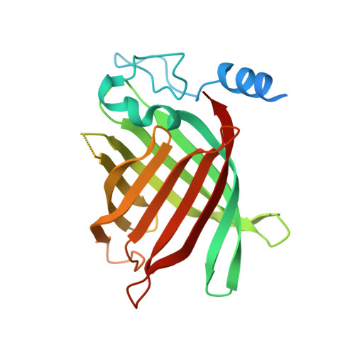
The Crystal Structure of Rv0813c from Mycobacterium tuberculosis Reveals a New Family of Fatty Acid-Binding Protein-Like Proteins in Bacteria

Shepard, W.Haouz, A.Grana, M.Buschiazzo, A.Betton, J.M.Cole, S.T.Alzari, P.M.

(2007) J Bacteriol 189: 1899-1904

  • DOI: https://doi.org/10.1128/JB.01435-06
  • Primary Citation of Related Structures:  
    2FWV

  • PubMed Abstract: 

    The gene Rv0813c from Mycobacterium tuberculosis, which codes for a hypothetical protein of unknown function, is conserved within the order Actinomycetales but absent elsewhere. The crystal structure of Rv0813c reveals a new family of proteins that resemble the fatty acid-binding proteins (FABPs) found in eukaryotes. Rv0813c adopts the 10-stranded beta-barrel fold typical of FABPs but lacks the double-helix insert that covers the entry to the binding site in the eukaryotic proteins. The barrel encloses a deep cavity, at the bottom of which a small cyclic ligand was found to bind to the hydroxyl group of Tyr192. This residue is part of a conserved Arg-X-Tyr motif much like the triad that binds the carboxylate group of fatty acids in FABPs. Most of the residues forming the internal surface of the cavity are conserved in homologous protein sequences found in CG-rich prokaryotes, strongly suggesting that Rv0813c is a member of a new family of bacterial FABP-like proteins that may have roles in the recognition, transport, and/or storage of small molecules in the bacterial cytosol.


  • Organizational Affiliation

    Unité de Biochimie Structurale, CNRS URA 2185, Institut Pasteur, 25 rue du Docteur Roux, 75724 Paris, France.


Macromolecules
Find similar proteins by:  (by identity cutoff)  |  3D Structure
Entity ID: 1
MoleculeChains Sequence LengthOrganismDetailsImage
hypothetical protein MtubF_01000852226Mycobacterium tuberculosis H37RvMutation(s): 0 
Gene Names: rv0813c
UniProt
Find proteins for P9WFG9 (Mycobacterium tuberculosis (strain ATCC 25618 / H37Rv))
Explore P9WFG9 
Go to UniProtKB:  P9WFG9
Entity Groups  
Sequence Clusters30% Identity50% Identity70% Identity90% Identity95% Identity100% Identity
UniProt GroupP9WFG9
Sequence Annotations
Expand
  • Reference Sequence
Experimental Data & Validation

Experimental Data

  • Method: X-RAY DIFFRACTION
  • Resolution: 1.70 Å
  • R-Value Free: 0.223 
  • R-Value Work: 0.181 
  • R-Value Observed: 0.183 
  • Space Group: I 41 2 2
Unit Cell:
Length ( Å )Angle ( ˚ )
a = 65.836α = 90
b = 65.836β = 90
c = 239.714γ = 90
Software Package:
Software NamePurpose
SCALAdata scaling
SOLVEphasing
RESOLVEphasing
REFMACrefinement
PDB_EXTRACTdata extraction
MOSFLMdata reduction
CCP4data scaling

Structure Validation

View Full Validation Report


Entry History 

Deposition Data

Revision History  (Full details and data files)

  • Version 1.0: 2006-08-03
    Type: Initial release
  • Version 1.1: 2008-05-01
    Changes: Version format compliance
  • Version 1.2: 2011-07-13
    Changes: Derived calculations, Source and taxonomy, Version format compliance
  • Version 1.3: 2013-06-26
    Changes: Derived calculations
  • Version 1.4: 2017-09-20
    Changes: Refinement description
  • Version 1.5: 2024-03-13
    Changes: Data collection, Database references, Derived calculations