


The electron transfer complex between cytochrome c552 and the CuA domain of the Thermus thermophilus ba3 oxidase - a combined NMR and computational approach
Muresanu, L., Pristovsek, P., Loehr, F., Maneg, O., Mukrasch, M.D., Rueterjans, H., Ludwig, B., Luecke, C.(2006) J Biol Chem 281: 14503-14513
- PubMed: 16554303
- DOI: https://doi.org/10.1074/jbc.M601108200
- Primary Citation of Related Structures:
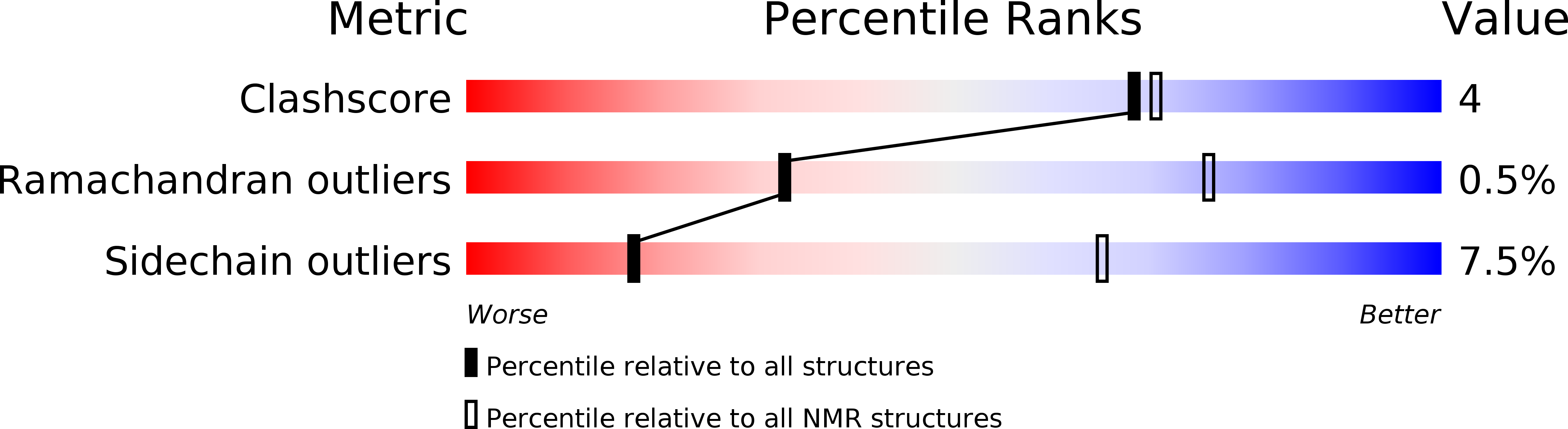
2FWL - PubMed Abstract:
The structural analysis of the redox complex between the soluble cytochrome c552 and the membrane-integral cytochrome ba3 oxidase of Thermus thermophilus is complicated by the transient nature of this protein-protein interaction. Using NMR-based chemical shift perturbation mapping, however, we identified the contact regions between cytochrome c552 and the CuA domain, the fully functional water-soluble fragment of subunit II of the ba3 oxidase. First we determined the complete backbone resonance assignments of both proteins for each redox state. Subsequently, two-dimensional [15N,1H]TROSY spectra recorded for each redox partner both in free and complexed state indicated those surface residues affected by complex formation between the two proteins. This chemical shift analysis performed for both redox states provided a topological description of the contact surface on each partner molecule. Remarkably, very pronounced indirect effects, which were observed on the back side of the heme cleft only in the reduced state, suggested that alterations of the electron distribution in the porphyrin ring due to formation of the protein-protein complex are apparently sensed even beyond the heme propionate groups. The contact residues of each redox partner, as derived from the chemical shift perturbation mapping, were employed for a protein-protein docking calculation that provided a structure ensemble of 10 closely related conformers representing the complex between cytochrome c552 and the CuA domain. Based on these structures, the electron transfer pathway from the heme of cytochrome c552 to the CuA center of the ba3 oxidase has been predicted.
Organizational Affiliation:
Institute of Biophysical Chemistry, Center for Biomolecular Magnetic Resonance, J. W. Goethe-University, Marie-Curie-Strasse 9, D-60439 Frankfurt am Main, Germany.


