2FPL

RadA recombinase in complex with AMP-PNP and low concentration of K+


Experimental Data Snapshot

  • Method: X-RAY DIFFRACTION
  • Resolution: 2.30 Å
  • R-Value Free: 0.249 
  • R-Value Work: 0.197 
  • R-Value Observed: 0.202 

wwPDB Validation   3D Report Full Report


Ligand Structure Quality Assessment 


This is version 1.5 of the entry. See complete history


Literature

Crystal Structure of Methanococcus Voltae Rada in Complex with Adp: hydrolysis-induced conformational change

Qian, X.Wu, Y.He, Y.Moya, I.A.Luo, Y.

(2005) Biochemistry 44: 13753-13761

  • DOI: https://doi.org/10.1021/bi051222i
  • Primary Citation of Related Structures:  
    2FPK, 2FPL, 2FPM

  • PubMed Abstract: 

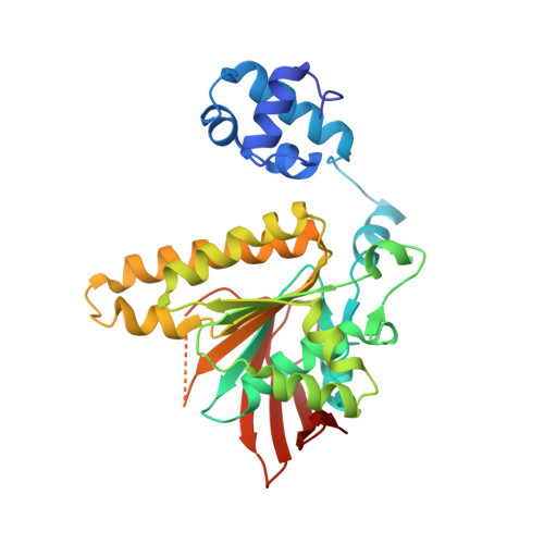
    Members of a superfamily of RecA-like recombinases facilitate a central strand exchange reaction in the DNA repair process. Archaeal RadA and Rad51 and eukaryal Rad51 and meiosis-specific DMC1 form a closely related group of recombinases distinct from bacterial RecA. Nevertheless, all such recombinases share a conserved core domain which carries the ATPase site and putative DNA-binding sites. Here we present the crystal structure of an archaeal RadA from Methanococcus voltae (MvRadA) in complex with ADP and Mg2+ at 2.1 A resolution. The crystallized RadA-ADP filament has an extended helical pitch similar to those of previously determined structures in the presence of nonhydrolyzable ATP analogue AMP-PNP. Structural comparison reveals two recurrent conformations with an extensive allosteric effect spanning the ATPase site and the putative DNA-binding L2 region. Varied conformations of the L2 region also imply a dynamic nature of recombinase-bound DNA.


  • Organizational Affiliation

    Department of Biochemistry, University of Saskatchewan, A3 Health Sciences Building, 107 Wiggins Road, Saskatoon, Saskatchewan, Canada S7N 5E5.


Macromolecules
Find similar proteins by:  (by identity cutoff)  |  3D Structure
Entity ID: 1
MoleculeChains Sequence LengthOrganismDetailsImage
DNA repair and recombination protein radA322Methanococcus voltaeMutation(s): 1 
Gene Names: RADA
UniProt
Find proteins for O73948 (Methanococcus voltae)
Explore O73948 
Go to UniProtKB:  O73948
Entity Groups  
Sequence Clusters30% Identity50% Identity70% Identity90% Identity95% Identity100% Identity
UniProt GroupO73948
Sequence Annotations
Expand
  • Reference Sequence
Experimental Data & Validation

Experimental Data

  • Method: X-RAY DIFFRACTION
  • Resolution: 2.30 Å
  • R-Value Free: 0.249 
  • R-Value Work: 0.197 
  • R-Value Observed: 0.202 
  • Space Group: P 61
Unit Cell:
Length ( Å )Angle ( ˚ )
a = 83.454α = 90
b = 83.454β = 90
c = 106.825γ = 120
Software Package:
Software NamePurpose
SAINTPLUSdata collection
LSCALEdata reduction
AMoREphasing
CNSrefinement
SAINTdata reduction
LSCALEdata scaling

Structure Validation

View Full Validation Report


Ligand Structure Quality Assessment 


Entry History 

Deposition Data

Revision History  (Full details and data files)

  • Version 1.0: 2006-01-31
    Type: Initial release
  • Version 1.1: 2008-05-01
    Changes: Version format compliance
  • Version 1.2: 2011-07-13
    Changes: Version format compliance
  • Version 1.3: 2017-10-18
    Changes: Refinement description
  • Version 1.4: 2021-10-20
    Changes: Database references, Derived calculations
  • Version 1.5: 2023-08-30
    Changes: Data collection, Refinement description