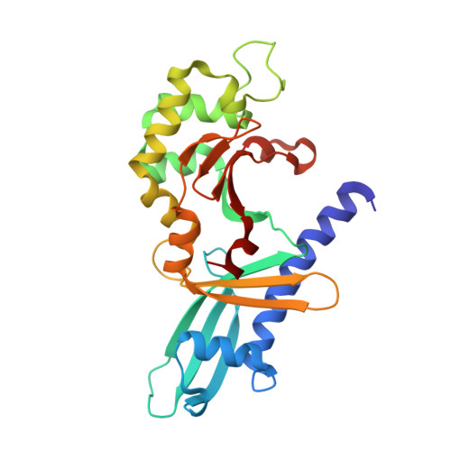
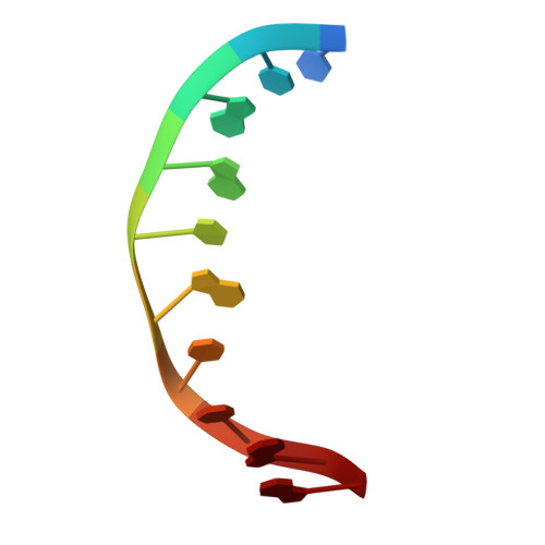
2FL3

Binary Complex of Restriction Endonuclease HinP1I with Cognate DNA


Experimental Data Snapshot

  • Method: X-RAY DIFFRACTION
  • Resolution: 2.39 Å
  • R-Value Free: 0.284 
  • R-Value Work: 0.246 
  • R-Value Observed: 0.249 

wwPDB Validation   3D Report Full Report


This is version 1.5 of the entry. See complete history


Literature

DNA nicking by HinP1I endonuclease: bending, base flipping and minor groove expansion.

Horton, J.R.Zhang, X.Maunus, R.Yang, Z.Wilson, G.G.Roberts, R.J.Cheng, X.

(2006) Nucleic Acids Res 34: 939-948

  • DOI: https://doi.org/10.1093/nar/gkj484
  • Primary Citation of Related Structures:  
    2FKC, 2FKH, 2FL3, 2FLC

  • PubMed Abstract: 

    HinP1I recognizes and cleaves the palindromic tetranucleotide sequence G downward arrowCGC in DNA. We report three structures of HinP1I-DNA complexes: in the presence of Ca(2+) (pre-reactive complex), in the absence of metal ion (binary complex) and in the presence of Mg(2+) (post-reactive complex). HinP1I forms a back-to-back dimer with two active sites and two DNA duplexes bound on the outer surfaces of the dimer facing away from each other. The 10 bp DNA duplexes undergo protein-induced distortions exhibiting features of A-, B- and Z-conformations: bending on one side (by intercalation of a phenylalanine side chain into the major groove), base flipping on the other side of the recognition site (by expanding the step rise distance of the local base pair to Z-form) and a local A-form conformation between the two central C:G base pairs of the recognition site (by binding of the N-terminal helix in the minor groove). In the pre- and post-reactive complexes, two metals (Ca(2+) or Mg(2+)) are found in the active site. The enzyme appears to cleave DNA sequentially, hydrolyzing first one DNA strand, as seen in the post-reactive complex in the crystalline state, and then the other, as supported by the observation that, in solution, a nicked DNA intermediate accumulates before linearization.


  • Organizational Affiliation

    Department of Biochemistry, Emory University School of Medicine 1510 Clifton Road, Atlanta, GA 30322, USA.


Macromolecules

Find similar proteins by:  (by identity cutoff)  |  3D Structure
Entity ID: 2
MoleculeChains Sequence LengthOrganismDetailsImage
R.HinP1I Restriction EndonucleaseC [auth A]247Haemophilus influenzaeMutation(s): 0 
Gene Names: hinP1IR
EC: 3.1.21.4
UniProt
Find proteins for Q5I6E6 (Haemophilus influenzae)
Explore Q5I6E6 
Go to UniProtKB:  Q5I6E6
Entity Groups  
Sequence Clusters30% Identity50% Identity70% Identity90% Identity95% Identity100% Identity
UniProt GroupQ5I6E6
Sequence Annotations
Expand
  • Reference Sequence

Find similar nucleic acids by:  Sequence   |   3D Structure  

Entity ID: 1
MoleculeChains LengthOrganismImage
5'-D(*CP*CP*AP*GP*CP*GP*CP*TP*GP*G)-3'A [auth C],
B [auth D]
10N/A
Sequence Annotations
Expand
  • Reference Sequence
Experimental Data & Validation

Experimental Data

  • Method: X-RAY DIFFRACTION
  • Resolution: 2.39 Å
  • R-Value Free: 0.284 
  • R-Value Work: 0.246 
  • R-Value Observed: 0.249 
  • Space Group: P 65 2 2
Unit Cell:
Length ( Å )Angle ( ˚ )
a = 100.855α = 90
b = 100.855β = 90
c = 139.406γ = 120
Software Package:
Software NamePurpose
MAR345data collection
SCALEPACKdata scaling
GLRFphasing
CNSrefinement

Structure Validation

View Full Validation Report


Entry History 

Deposition Data

  • Released Date: 2006-02-21 
  • Deposition Author(s): Horton, J.R.

Revision History  (Full details and data files)

  • Version 1.0: 2006-02-21
    Type: Initial release
  • Version 1.1: 2008-05-01
    Changes: Version format compliance
  • Version 1.2: 2011-07-13
    Changes: Version format compliance
  • Version 1.3: 2017-10-18
    Changes: Refinement description
  • Version 1.4: 2018-01-31
    Changes: Database references
  • Version 1.5: 2023-08-30
    Changes: Data collection, Database references, Refinement description