2FDC

Structural Basis of DNA Damage Recognition and Processing by UvrB: crystal structure of a UvrB/DNA complex


Experimental Data Snapshot

  • Method: X-RAY DIFFRACTION
  • Resolution: 3.30 Å
  • R-Value Free: 0.258 
  • R-Value Work: 0.205 
  • R-Value Observed: 0.210 

wwPDB Validation   3D Report Full Report


Ligand Structure Quality Assessment 


This is version 1.4 of the entry. See complete history


Literature

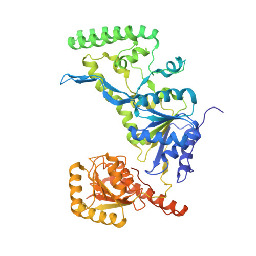
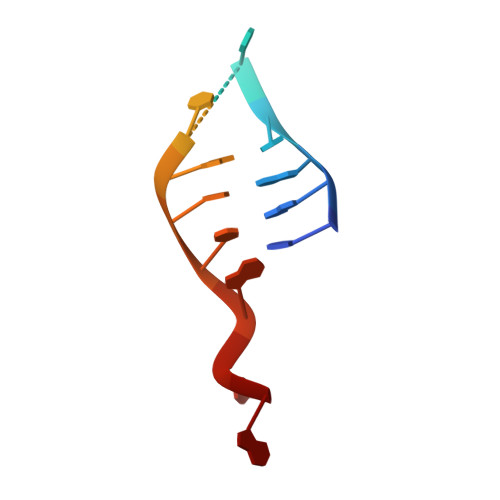
Structural basis for DNA recognition and processing by UvrB.

Truglio, J.J.Karakas, E.Rhau, B.Wang, H.DellaVecchia, M.J.Van Houten, B.Kisker, C.

(2006) Nat Struct Mol Biol 13: 360-364

  • DOI: https://doi.org/10.1038/nsmb1072
  • Primary Citation of Related Structures:  
    2FDC

  • PubMed Abstract: 

    DNA-damage recognition in the nucleotide excision repair (NER) cascade is a complex process, operating on a wide variety of damages. UvrB is the central component in prokaryotic NER, directly involved in DNA-damage recognition and guiding the DNA through repair synthesis. We report the first structure of a UvrB-double-stranded DNA complex, providing insights into the mechanism by which UvrB binds DNA, leading to formation of the preincision complex. One DNA strand, containing a 3' overhang, threads behind a beta-hairpin motif of UvrB, indicating that this motif inserts between the strands of the double helix, thereby locking down either the damaged or undamaged strand. The nucleotide directly behind the beta-hairpin is flipped out and inserted into a small, highly conserved pocket in UvrB.


  • Organizational Affiliation

    Department of Pharmacological Sciences, Stony Brook University, Stony Brook, New York 11794-5115, USA.


Macromolecules

Find similar proteins by:  (by identity cutoff)  |  3D Structure
Entity ID: 2
MoleculeChains Sequence LengthOrganismDetailsImage
UvrABC system protein BC [auth A],
D [auth B]
658[Bacillus] caldotenaxMutation(s): 0 
Gene Names: uvrB
UniProt
Find proteins for P56981 (Bacillus caldotenax)
Explore P56981 
Go to UniProtKB:  P56981
Entity Groups  
Sequence Clusters30% Identity50% Identity70% Identity90% Identity95% Identity100% Identity
UniProt GroupP56981
Sequence Annotations
Expand
  • Reference Sequence

Find similar nucleic acids by:  Sequence   |   3D Structure  

Entity ID: 1
MoleculeChains LengthOrganismImage
5'-D(P*CP*GP*GP*CP*TP*CP*CP*AP*TP*CP*TP*CP*TP*AP*CP*CP*GP*CP*AP*A)-3'A [auth C],
B [auth D]
20N/A
Sequence Annotations
Expand
  • Reference Sequence
Small Molecules
Ligands 1 Unique
IDChains Name / Formula / InChI Key2D Diagram3D Interactions
FLQ
Query on FLQ

Download Ideal Coordinates CCD File 
E [auth C]N-[6-(ACETYLAMINO)HEXYL]-3',6'-DIHYDROXY-3-OXO-3H-SPIRO[2-BENZOFURAN-1,9'-XANTHENE]-6-CARBOXAMIDE
C29 H28 N2 O7
NYDPRVGMTFCAQC-UHFFFAOYSA-N
Experimental Data & Validation

Experimental Data

  • Method: X-RAY DIFFRACTION
  • Resolution: 3.30 Å
  • R-Value Free: 0.258 
  • R-Value Work: 0.205 
  • R-Value Observed: 0.210 
  • Space Group: P 32 2 1
Unit Cell:
Length ( Å )Angle ( ˚ )
a = 153.265α = 90
b = 153.265β = 90
c = 160.17γ = 120
Software Package:
Software NamePurpose
DENZOdata reduction
SCALEPACKdata scaling
REFMACrefinement
PDB_EXTRACTdata extraction
HKL-2000data reduction
AMoREphasing

Structure Validation

View Full Validation Report


Ligand Structure Quality Assessment 


Entry History 

Deposition Data

Revision History  (Full details and data files)

  • Version 1.0: 2006-03-14
    Type: Initial release
  • Version 1.1: 2008-05-01
    Changes: Version format compliance
  • Version 1.2: 2011-07-13
    Changes: Advisory, Version format compliance
  • Version 1.3: 2017-10-18
    Changes: Refinement description
  • Version 1.4: 2024-02-14
    Changes: Data collection, Database references, Derived calculations