2F62

Crystal structure of Nucleoside 2-deoxyribosyltransferase from Trypanosoma brucei at 1.5 A resolution with (2-ETHYLPHENYL)METHANOL bound


Experimental Data Snapshot

  • Method: X-RAY DIFFRACTION
  • Resolution: 1.50 Å
  • R-Value Free: 0.186 
  • R-Value Work: 0.167 
  • R-Value Observed: 0.168 

wwPDB Validation   3D Report Full Report


This is version 1.4 of the entry. See complete history


Literature

Using fragment cocktail crystallography to assist inhibitor design of Trypanosoma brucei nucleoside 2-deoxyribosyltransferase.

Bosch, J.Robien, M.A.Mehlin, C.Boni, E.Riechers, A.Buckner, F.S.Van Voorhis, W.C.Myler, P.J.Worthey, E.A.DeTitta, G.Luft, J.R.Lauricella, A.Gulde, S.Anderson, L.A.Kalyuzhniy, O.Neely, H.M.Ross, J.Earnest, T.N.Soltis, M.Schoenfeld, L.Zucker, F.Merritt, E.A.Fan, E.Verlinde, C.L.Hol, W.G.J.

(2006) J Med Chem 49: 5939-5946

  • DOI: https://doi.org/10.1021/jm060429m
  • Primary Citation of Related Structures:  
    2A0K, 2F2T, 2F62, 2F64, 2F67

  • PubMed Abstract: 

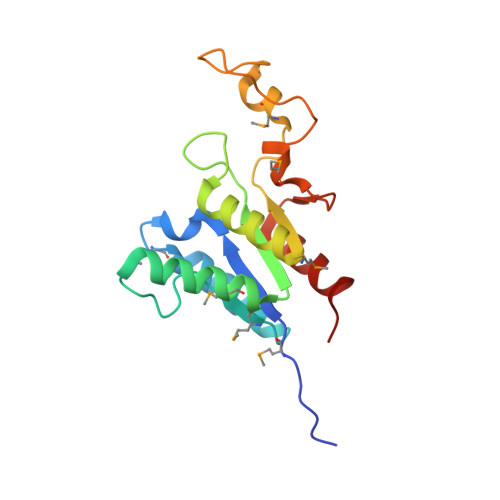
    The 1.8 A resolution de novo structure of nucleoside 2-deoxyribosyltransferase (EC 2.4.2.6) from Trypanosoma brucei (TbNDRT) has been determined by SADa phasing in an unliganded state and several ligand-bound states. This enzyme is important in the salvage pathway of nucleoside recycling. To identify novel lead compounds, we exploited "fragment cocktail soaks". Out of 304 compounds tried in 31 cocktails, four compounds could be identified crystallographically in the active site. In addition, we demonstrated that very short soaks of approximately 10 s are sufficient even for rather hydrophobic ligands to bind in the active site groove, which is promising for the application of similar soaking experiments to less robust crystals of other proteins.


  • Organizational Affiliation

    Department of Biochemistry, Division of Infectious Disease, and Howard Hughes Medical Institute, University of Washington, Seattle, Washington 98195, USA.


Macromolecules
Find similar proteins by:  (by identity cutoff)  |  3D Structure
Entity ID: 1
MoleculeChains Sequence LengthOrganismDetailsImage
Nucleoside 2-deoxyribosyltransferase
A, B
161Trypanosoma bruceiMutation(s): 7 
Gene Names: Tb05.30H13.400
EC: 2.4.2.6
UniProt
Find proteins for Q57VC7 (Trypanosoma brucei brucei (strain 927/4 GUTat10.1))
Explore Q57VC7 
Go to UniProtKB:  Q57VC7
Entity Groups  
Sequence Clusters30% Identity50% Identity70% Identity90% Identity95% Identity100% Identity
UniProt GroupQ57VC7
Sequence Annotations
Expand
  • Reference Sequence
Small Molecules
Modified Residues  1 Unique
IDChains TypeFormula2D DiagramParent
MSE
Query on MSE
A, B
L-PEPTIDE LINKINGC5 H11 N O2 SeMET
Experimental Data & Validation

Experimental Data

  • Method: X-RAY DIFFRACTION
  • Resolution: 1.50 Å
  • R-Value Free: 0.186 
  • R-Value Work: 0.167 
  • R-Value Observed: 0.168 
  • Space Group: C 1 2 1
Unit Cell:
Length ( Å )Angle ( ˚ )
a = 58.614α = 90
b = 75.913β = 90.03
c = 85.957γ = 90
Software Package:
Software NamePurpose
REFMACrefinement
MOSFLMdata reduction
CCP4data scaling
MOLREPphasing

Structure Validation

View Full Validation Report


Entry History 

Deposition Data

Revision History  (Full details and data files)

  • Version 1.0: 2005-12-06
    Type: Initial release
  • Version 1.1: 2008-05-01
    Changes: Version format compliance
  • Version 1.2: 2011-07-13
    Changes: Advisory, Version format compliance
  • Version 1.3: 2023-08-23
    Changes: Data collection, Database references, Derived calculations, Refinement description
  • Version 1.4: 2023-11-15
    Changes: Data collection