2F3N

Crystal Structure of the native Shank SAM domain.


Experimental Data Snapshot

  • Method: X-RAY DIFFRACTION
  • Resolution: 2.10 Å
  • R-Value Free: 0.251 
  • R-Value Work: 0.206 
  • R-Value Observed: 0.206 

wwPDB Validation   3D Report Full Report


This is version 1.4 of the entry. See complete history


Literature

An architectural framework that may lie at the core of the postsynaptic density.

Baron, M.K.Boeckers, T.M.Vaida, B.Faham, S.Gingery, M.Sawaya, M.R.Salyer, D.Gundelfinger, E.D.Bowie, J.U.

(2006) Science 311: 531-535

  • DOI: https://doi.org/10.1126/science.1118995
  • Primary Citation of Related Structures:  
    2F3N, 2F44

  • PubMed Abstract: 

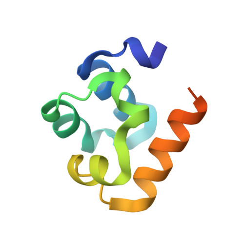
    The postsynaptic density (PSD) is a complex assembly of proteins associated with the postsynaptic membrane that organizes neurotransmitter receptors, signaling pathways, and regulatory elements within a cytoskeletal matrix. Here we show that the sterile alpha motif domain of rat Shank3/ProSAP2, a master scaffolding protein located deep within the PSD, can form large sheets composed of helical fibers stacked side by side. Zn2+, which is found in high concentrations in the PSD, binds tightly to Shank3 and may regulate assembly. Sheets of the Shank protein could form a platform for the construction of the PSD complex.


  • Organizational Affiliation

    Department of Chemistry and Biochemistry, Molecular Biology Institute, University of California, Los Angeles, 611 Charles E. Young Drive East, Los Angeles, CA 90095-1570, USA.


Macromolecules
Find similar proteins by:  (by identity cutoff)  |  3D Structure
Entity ID: 1
MoleculeChains Sequence LengthOrganismDetailsImage
SH3 and multiple ankyrin repeat domains 3
A, B, C
76Rattus norvegicusMutation(s): 1 
Gene Names: Shank3
UniProt
Find proteins for Q9JLU4 (Rattus norvegicus)
Explore Q9JLU4 
Go to UniProtKB:  Q9JLU4
Entity Groups  
Sequence Clusters30% Identity50% Identity70% Identity90% Identity95% Identity100% Identity
UniProt GroupQ9JLU4
Sequence Annotations
Expand
  • Reference Sequence
Experimental Data & Validation

Experimental Data

  • Method: X-RAY DIFFRACTION
  • Resolution: 2.10 Å
  • R-Value Free: 0.251 
  • R-Value Work: 0.206 
  • R-Value Observed: 0.206 
  • Space Group: P 65
Unit Cell:
Length ( Å )Angle ( ˚ )
a = 104.715α = 90
b = 104.715β = 90
c = 40.188γ = 120
Software Package:
Software NamePurpose
DENZOdata reduction
SCALEPACKdata scaling
MLPHAREphasing
CNSrefinement

Structure Validation

View Full Validation Report


Entry History 

Deposition Data

Revision History  (Full details and data files)

  • Version 1.0: 2006-02-07
    Type: Initial release
  • Version 1.1: 2008-05-01
    Changes: Version format compliance
  • Version 1.2: 2011-07-13
    Changes: Version format compliance
  • Version 1.3: 2021-10-20
    Changes: Database references
  • Version 1.4: 2024-02-14
    Changes: Data collection