2EWS

Crystal structure of S.aureus pantothenate kinase


Experimental Data Snapshot

  • Method: X-RAY DIFFRACTION
  • Resolution: 2.05 Å
  • R-Value Free: 0.289 
  • R-Value Work: 0.216 
  • R-Value Observed: 0.219 

wwPDB Validation   3D Report Full Report


Ligand Structure Quality Assessment 


This is version 1.3 of the entry. See complete history


Literature

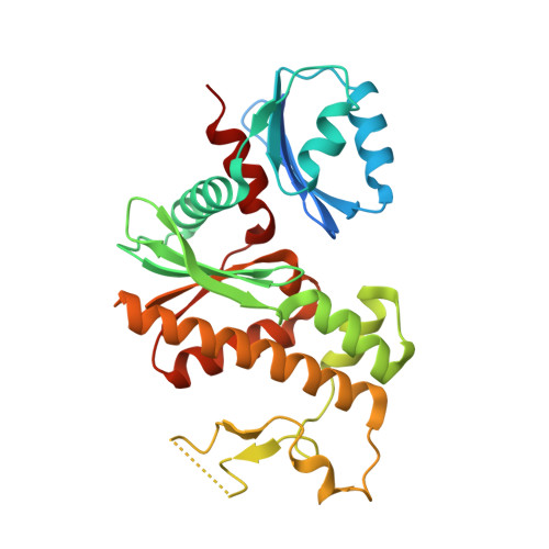
Prokaryotic Type II and Type III Pantothenate Kinases: The Same Monomer Fold Creates Dimers with Distinct Catalytic Properties.

Hong, B.S.Yun, M.K.Zhang, Y.M.Chohnan, S.Rock, C.O.White, S.W.Jackowski, S.Park, H.W.Leonardi, R.

(2006) Structure 14: 1251-1261

  • DOI: https://doi.org/10.1016/j.str.2006.06.008
  • Primary Citation of Related Structures:  
    2EWS, 2F9T, 2F9W

  • PubMed Abstract: 

    Three distinct isoforms of pantothenate kinase (CoaA) in bacteria catalyze the first step in coenzyme A biosynthesis. The structures of the type II (Staphylococcus aureus, SaCoaA) and type III (Pseudomonas aeruginosa, PaCoaA) enzymes reveal that they assemble nearly identical subunits with actin-like folds into dimers that exhibit distinct biochemical properties. PaCoaA has a fully enclosed pantothenate binding pocket and requires a monovalent cation to weakly bind ATP in an open cavity that does not interact with the adenine nucleotide. Pantothenate binds to an open pocket in SaCoaA that strongly binds ATP by using a classical P loop architecture coupled with specific interactions with the adenine moiety. The PaCoaA*Pan binary complex explains the resistance of bacteria possessing this isoform to the pantothenamide antibiotics, and the similarity between SaCoaA and human pantothenate kinase 2 explains the molecular basis for the development of the neurodegenerative phenotype in three mutations in the human protein.


  • Organizational Affiliation

    Structural Genomics Consortium, University of Toronto, Toronto, Ontario M5G 1L5, Canada.


Macromolecules
Find similar proteins by:  (by identity cutoff)  |  3D Structure
Entity ID: 1
MoleculeChains Sequence LengthOrganismDetailsImage
Pantothenate kinase
A, B
287Staphylococcus aureus subsp. aureus MSSA476Mutation(s): 0 
EC: 2.7.1.33
UniProt
Find proteins for Q6G7I0 (Staphylococcus aureus (strain MSSA476))
Explore Q6G7I0 
Go to UniProtKB:  Q6G7I0
Entity Groups  
Sequence Clusters30% Identity50% Identity70% Identity90% Identity95% Identity100% Identity
UniProt GroupQ6G7I0
Sequence Annotations
Expand
  • Reference Sequence
Small Molecules
Binding Affinity Annotations 
IDSourceBinding Affinity
ANP PDBBind:  2EWS Kd: 240 (nM) from 1 assay(s)
Experimental Data & Validation

Experimental Data

  • Method: X-RAY DIFFRACTION
  • Resolution: 2.05 Å
  • R-Value Free: 0.289 
  • R-Value Work: 0.216 
  • R-Value Observed: 0.219 
  • Space Group: P 63
Unit Cell:
Length ( Å )Angle ( ˚ )
a = 104.044α = 90
b = 104.044β = 90
c = 85.718γ = 120
Software Package:
Software NamePurpose
REFMACrefinement
HKL-2000data reduction
SCALEPACKdata scaling
CNSphasing
MOLREPphasing

Structure Validation

View Full Validation Report


Ligand Structure Quality Assessment 


Entry History 

Deposition Data

Revision History  (Full details and data files)

  • Version 1.0: 2006-09-12
    Type: Initial release
  • Version 1.1: 2008-05-01
    Changes: Version format compliance
  • Version 1.2: 2011-07-13
    Changes: Source and taxonomy, Version format compliance
  • Version 1.3: 2024-02-14
    Changes: Data collection, Database references, Derived calculations