High resolution x-ray analysis of two mutants of a curaremimetic snake toxin
Gaucher, J.F., Menez, R., Arnoux, B., Menez, A., Ducruix, A.(2000) Eur J Biochem 267: 1323-1329
- PubMed: 10691969
- DOI: https://doi.org/10.1046/j.1432-1327.2000.01099.x
- Primary Citation of Related Structures:
2ERA, 3ERA - PubMed Abstract:
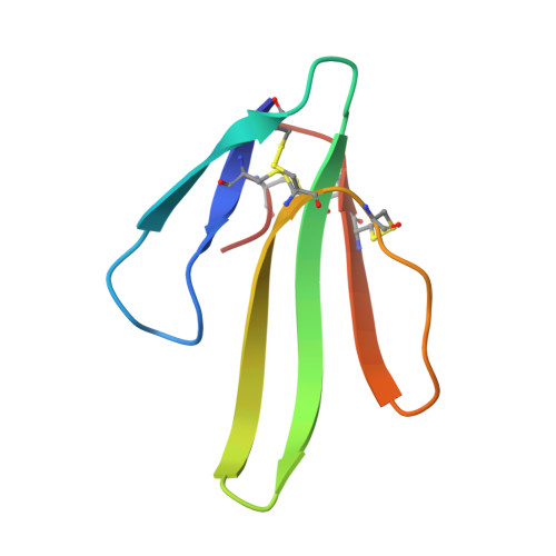
A previous mutational analysis of erabutoxin a (Ea), a curaremimetic toxin from sea snake venom, showed that the substitutions S8G and S8T caused, respectively, 176-fold and 780-fold affinity decreases for the nicotinic acetylcholine receptor (AchR). In view of the fact that the side-chain of Ser8 is buried in the wild-type toxin, we wondered whether these affinity changes reflect a direct binding contribution of S8 to the receptor and/or conformational changes that could have occurred in Ea as a result of the introduced mutations. To approach this question, we solved X-ray structures of the two mutants S8G and S8T at high resolution (0.18 nm and 0.17 nm, with R factors of 18.0% and 17.9%, respectively). The data show that none of the mutations significantly modified the toxin structure. Even within the site where the toxin binds to the receptor the backbone conformation remained unchanged. Therefore, the low affinities of the mutants S8T and S8G cannot be explained by a large conformational change of the toxin structure. Although we cannot exclude the possibility that undetectable structural changes have occurred in the toxin mutants, our data support the view that, although buried between loop I and II, S8 is part of the functional epitope of the toxin.
Organizational Affiliation:
Laboratoire d'Enzymologie et de Biochimie Structurales, UPR 9063 CNRS, 91198 Gif-sur-Yvette, France.