


Crystal structure of Human RNA-Binding Protein 12
Rehse, P.H., Nakano, N., Yokoyama, S.To be published.
Experimental Data Snapshot
wwPDB Validation 3D Report Full Report

Entity ID: 1 | |||||
|---|---|---|---|---|---|
| Molecule | Chains | Sequence Length | Organism | Details | Image |
| RNA-binding protein 12 | 95 | Homo sapiens | Mutation(s): 0 | 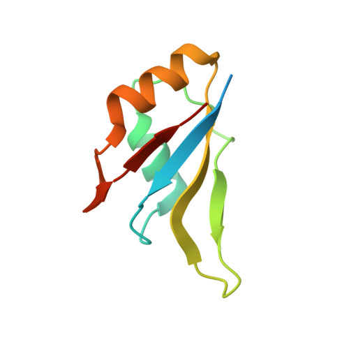 | |
UniProt & NIH Common Fund Data Resources | |||||
Find proteins for Q9NTZ6 (Homo sapiens) Explore Q9NTZ6 Go to UniProtKB: Q9NTZ6 | |||||
PHAROS: Q9NTZ6 GTEx: ENSG00000244462 | |||||
Entity Groups | |||||
| Sequence Clusters | 30% Identity50% Identity70% Identity90% Identity95% Identity100% Identity | ||||
| UniProt Group | Q9NTZ6 | ||||
Sequence AnnotationsExpand | |||||
| |||||
| Length ( Å ) | Angle ( ˚ ) |
|---|---|
| a = 107.02 | α = 90 |
| b = 107.02 | β = 90 |
| c = 58.89 | γ = 120 |
| Software Name | Purpose |
|---|---|
| DENZO | data reduction |
| SCALEPACK | data scaling |
| MOLREP | phasing |
| CNS | refinement |
| PDB_EXTRACT | data extraction |
| ADSC | data collection |
| HKL-2000 | data reduction |
RCSB PDB (citation) is hosted by
RCSB PDB is a member of the

