2E85

Crystal Structure of the Hydrogenase 3 Maturation protease


Experimental Data Snapshot

  • Method: X-RAY DIFFRACTION
  • Resolution: 1.70 Å
  • R-Value Free: 0.258 
  • R-Value Work: 0.248 
  • R-Value Observed: 0.248 

wwPDB Validation   3D Report Full Report


This is version 1.2 of the entry. See complete history


Literature

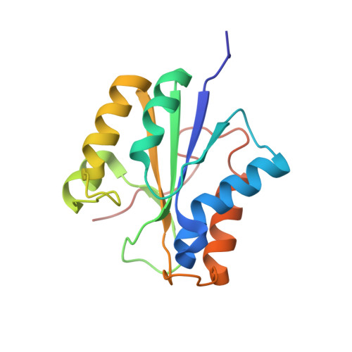
Crystal structure of hydrogenase maturating endopeptidase HycI from Escherichia coli

Kumarevel, T.Tanaka, T.Bessho, Y.Shinkai, A.Yokoyama, S.

(2009) Biochem Biophys Res Commun 389: 310-314

  • DOI: https://doi.org/10.1016/j.bbrc.2009.08.135
  • Primary Citation of Related Structures:  
    2E85

  • PubMed Abstract: 

    The maturation of [NiFe]-hydrogenases is a catalyzed process involving the activities of at least seven proteins. The last step consists of the endoproteolytic cleavage of the precursor of the large subunit, after the [NiFe]-metal center has been assembled. The HycI endopeptidase is involved in the C-terminal processing of HycE, the large subunit of hydrogenase 3 from Escherichia coli. Although HycI has been well characterized biochemically, the crystallization of the protein has been quite challenging. Here, we present the crystal structure of HycI at 1.70 A resolution. The crystal structure resembles the recently reported solution structure (NMR) of the same protein and the holo-HyPD structure of the same family, but a significant conformational change is observed at the L5 loop, as compared with the solution structures of HycI and HyPD. In our crystal structure, three specific metal binding sites (Ca1-3) were identified and these metal ions are possibly involved in the C-terminal cleavage of HycE.


  • Organizational Affiliation

    RIKEN SPring-8 Center, Harima Institute, 1-1-1 Kouto, Sayo, Hyogo 679-5148, Japan. tskvel@spring8.or.jp


Macromolecules
Find similar proteins by:  (by identity cutoff)  |  3D Structure
Entity ID: 1
MoleculeChains Sequence LengthOrganismDetailsImage
Hydrogenase 3 maturation protease
A, B
159Escherichia coliMutation(s): 1 
EC: 3.4
UniProt
Find proteins for P0AEV9 (Escherichia coli (strain K12))
Explore P0AEV9 
Go to UniProtKB:  P0AEV9
Entity Groups  
Sequence Clusters30% Identity50% Identity70% Identity90% Identity95% Identity100% Identity
UniProt GroupP0AEV9
Sequence Annotations
Expand
  • Reference Sequence
Experimental Data & Validation

Experimental Data

  • Method: X-RAY DIFFRACTION
  • Resolution: 1.70 Å
  • R-Value Free: 0.258 
  • R-Value Work: 0.248 
  • R-Value Observed: 0.248 
  • Space Group: P 21 21 2
Unit Cell:
Length ( Å )Angle ( ˚ )
a = 87.425α = 90
b = 54.984β = 90
c = 67.964γ = 90
Software Package:
Software NamePurpose
CNSrefinement
ADSCdata collection
HKL-2000data reduction
HKL-2000data scaling
SOLVEphasing

Structure Validation

View Full Validation Report


Entry History 

Deposition Data

Revision History  (Full details and data files)

  • Version 1.0: 2008-01-22
    Type: Initial release
  • Version 1.1: 2011-07-13
    Changes: Version format compliance
  • Version 1.2: 2021-11-10
    Changes: Database references, Derived calculations