2E4Z

Crystal structure of the ligand-binding region of the group III metabotropic glutamate receptor


Experimental Data Snapshot

  • Method: X-RAY DIFFRACTION
  • Resolution: 3.30 Å
  • R-Value Free: 0.324 
  • R-Value Work: 0.270 
  • R-Value Observed: 0.274 

wwPDB Validation   3D Report Full Report


This is version 1.3 of the entry. See complete history


Literature

Structures of the extracellular regions of the group II/III metabotropic glutamate receptors

Muto, T.Tsuchiya, D.Morikawa, K.Jingami, H.

(2007) Proc Natl Acad Sci U S A 104: 3759-3764

  • DOI: https://doi.org/10.1073/pnas.0611577104
  • Primary Citation of Related Structures:  
    2E4U, 2E4V, 2E4W, 2E4X, 2E4Y, 2E4Z

  • PubMed Abstract: 

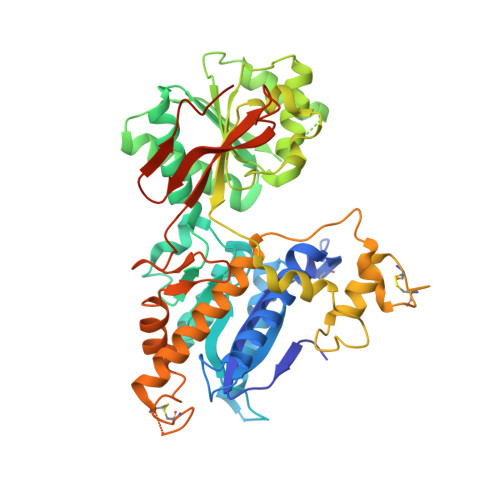
    Metabotropic glutamate receptors play major roles in the activation of excitatory synapses in the central nerve system. We determined the crystal structure of the entire extracellular region of the group II receptor and that of the ligand-binding region of the group III receptor. A comparison among groups I, II, and III provides the structural basis that could account for the discrimination of group-specific agonists. Furthermore, the structure of group II includes the cysteine-rich domain, which is tightly linked to the ligand-binding domain by a disulfide bridge, suggesting a potential role in transmitting a ligand-induced conformational change into the downstream transmembrane region. The structure also reveals the lateral interaction between the two cysteine-rich domains, which could stimulate clustering of the dimeric receptors on the cell surface. We propose a general activation mechanism of the dimeric receptor coupled with both ligand-binding and interprotomer rearrangements.


  • Organizational Affiliation

    Biomolecular Engineering Research Institute, 6-2-3 Furuedai, Suita, Osaka 565-0874, Japan.


Macromolecules
Find similar proteins by:  (by identity cutoff)  |  3D Structure
Entity ID: 1
MoleculeChains Sequence LengthOrganismDetailsImage
Metabotropic glutamate receptor 7501Rattus norvegicusMutation(s): 0 
UniProt
Find proteins for P35400 (Rattus norvegicus)
Explore P35400 
Go to UniProtKB:  P35400
Entity Groups  
Sequence Clusters30% Identity50% Identity70% Identity90% Identity95% Identity100% Identity
UniProt GroupP35400
Sequence Annotations
Expand
  • Reference Sequence
Small Molecules
Ligands 1 Unique
IDChains Name / Formula / InChI Key2D Diagram3D Interactions
MES
Query on MES

Download Ideal Coordinates CCD File 
B [auth A]2-(N-MORPHOLINO)-ETHANESULFONIC ACID
C6 H13 N O4 S
SXGZJKUKBWWHRA-UHFFFAOYSA-N
Experimental Data & Validation

Experimental Data

  • Method: X-RAY DIFFRACTION
  • Resolution: 3.30 Å
  • R-Value Free: 0.324 
  • R-Value Work: 0.270 
  • R-Value Observed: 0.274 
  • Space Group: P 31 2 1
Unit Cell:
Length ( Å )Angle ( ˚ )
a = 92.431α = 90
b = 92.431β = 90
c = 114.332γ = 120
Software Package:
Software NamePurpose
REFMACrefinement
ADSCdata collection
MOSFLMdata reduction
CCP4data scaling
AMoREphasing

Structure Validation

View Full Validation Report


Entry History 

Deposition Data

Revision History  (Full details and data files)

  • Version 1.0: 2007-02-27
    Type: Initial release
  • Version 1.1: 2008-04-30
    Changes: Version format compliance
  • Version 1.2: 2011-07-13
    Changes: Advisory, Version format compliance
  • Version 1.3: 2023-10-25
    Changes: Data collection, Database references, Derived calculations, Refinement description