Kinetic and structural analysis of enzyme sliding on a substrate: multiple attack in beta-amylase
Ishikawa, K., Nakatani, H., Katsuya, Y., Fukazawa, C.(2007) Biochemistry 46: 792-798
- PubMed: 17223700
- DOI: https://doi.org/10.1021/bi061605w
- Primary Citation of Related Structures:
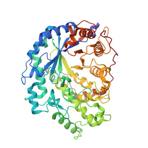
2DQX - PubMed Abstract:
Beta-amylase (EC 3.2.1.2) is starch-hydrolyzing exo-type enzyme that can catalyze the successive liberation of beta-maltose from the nonreducing ends of alpha-1,4-linked glucopyranosyl polymers. There is a well-known phenomenon called multiple or repetitive attack where the enzyme releases several maltose molecules in a single enzyme-substrate complex. In order to understand it further, we examined the beta-amylase-catalyzed reaction using maltooligosaccharides. The Monte Carlo method was applied for simulation of the beta-amylase-catalyzed reaction including the multiple attack mechanism. Through site-directed mutagenesis, we have successfully prepared a mutant enzyme which may be simulated as a multiple attack action reduced one with retaining significant hydrolytic activity. From the results of X-ray structure analysis of the mutant enzyme, it was clarified that one carboxyl residue plays a very important role in the multiple attack. The multiple attack action needs the force of enzyme sliding on the substrate. In addition, it is important for the multiple attack that the enzyme and substrate have the characteristics of a stable productive substrate-enzyme complex through a hydrogen bond between the nonreducing end of the substrate and the carboxyl residue of the enzyme.
Organizational Affiliation:
National Institute of Advanced Industrial Science and Technology, 1-8-31 Midorigaoka, Ikeda, Osaka 563-8577, Japan. kazu-ishikawa@aist.go.jp