2DPK

The Crystal Structure of the Primary Ca2+ Sensor of the Na+/Ca2+ Exchanger


Experimental Data Snapshot

  • Method: X-RAY DIFFRACTION
  • Resolution: 2.50 Å
  • R-Value Free: 0.284 
  • R-Value Work: 0.222 
  • R-Value Observed: 0.225 

wwPDB Validation   3D Report Full Report


This is version 1.2 of the entry. See complete history


Literature

The crystal structure of the primary Ca2+ sensor of the na+/ca2+ exchanger reveals a novel Ca2+ binding motif.

Nicoll, D.A.Sawaya, M.Kwon, S.Cascio, D.Philipson, K.D.Abramson, J.

(2006) J Biol Chem 281: 21577-21581

  • DOI: https://doi.org/10.1074/jbc.C600117200
  • Primary Citation of Related Structures:  
    2DPK

  • PubMed Abstract: 

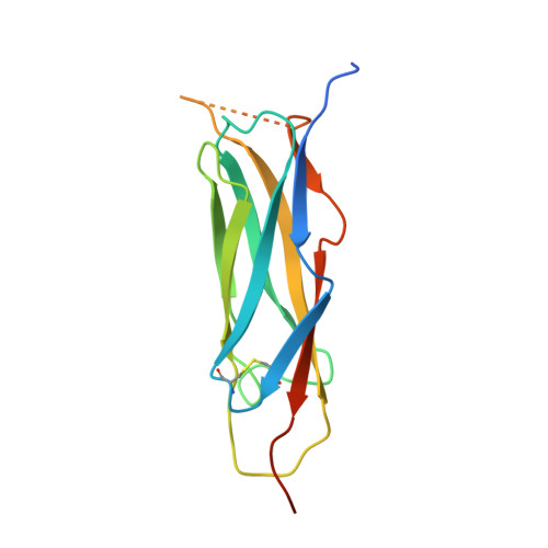
    The Na+/Ca2+ exchanger is a plasma membrane protein that regulates intracellular Ca2+ levels in cardiac myocytes. Transport activity is governed by Ca2+, and the primary Ca2+ sensor (CBD1) is located in a large cytoplasmic loop connecting two transmembrane helices. The binding of Ca2+ to the CBD1 sensory domain results in conformational changes that stimulate the exchanger to extrude Ca2+. Here, we present a crystal structure of CBD1 at 2.5A resolution, which reveals a novel Ca2+ binding site consisting of four Ca2+ ions arranged in a tight planar cluster. This intricate coordination pattern for a Ca2+ binding cluster is indicative of a highly sensitive Ca2+ sensor and may represent a general platform for Ca2+ sensing.


  • Organizational Affiliation

    Department of Physiology and the Cardiovascular Research Laboratories, David Geffen School of Medicine, University of California, Los Angeles, California 90095.


Macromolecules
Find similar proteins by:  (by identity cutoff)  |  3D Structure
Entity ID: 1
MoleculeChains Sequence LengthOrganismDetailsImage
Sodium/calcium exchanger 1152Canis lupus familiarisMutation(s): 0 
UniProt
Find proteins for P23685 (Canis lupus familiaris)
Explore P23685 
Go to UniProtKB:  P23685
Entity Groups  
Sequence Clusters30% Identity50% Identity70% Identity90% Identity95% Identity100% Identity
UniProt GroupP23685
Sequence Annotations
Expand
  • Reference Sequence
Experimental Data & Validation

Experimental Data

  • Method: X-RAY DIFFRACTION
  • Resolution: 2.50 Å
  • R-Value Free: 0.284 
  • R-Value Work: 0.222 
  • R-Value Observed: 0.225 
  • Space Group: P 21 21 2
Unit Cell:
Length ( Å )Angle ( ˚ )
a = 59.624α = 90
b = 45.524β = 90
c = 57.269γ = 90
Software Package:
Software NamePurpose
REFMACrefinement

Structure Validation

View Full Validation Report


Entry History 

Deposition Data

Revision History  (Full details and data files)

  • Version 1.0: 2006-06-13
    Type: Initial release
  • Version 1.1: 2008-04-30
    Changes: Version format compliance
  • Version 1.2: 2011-07-13
    Changes: Version format compliance