2DFV

Hyperthermophilic threonine dehydrogenase from Pyrococcus horikoshii


Experimental Data Snapshot

  • Method: X-RAY DIFFRACTION
  • Resolution: 2.05 Å
  • R-Value Free: 0.239 
  • R-Value Work: 0.204 
  • R-Value Observed: 0.206 

wwPDB Validation   3D Report Full Report


Ligand Structure Quality Assessment 


This is version 1.2 of the entry. See complete history


Literature

The first crystal structure of L-threonine dehydrogenase.

Ishikawa, K.Higashi, N.Nakamura, T.Matsuura, T.Nakagawa, A.

(2007) J Mol Biol 366: 857-867

  • DOI: https://doi.org/10.1016/j.jmb.2006.11.060
  • Primary Citation of Related Structures:  
    2DFV

  • PubMed Abstract: 

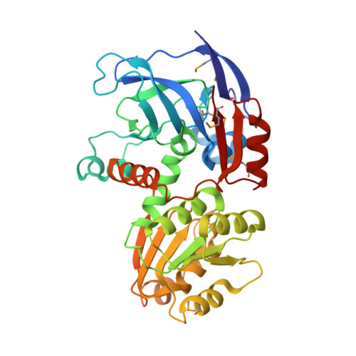
    L-threonine dehydrogenase (TDH) is an enzyme that catalyzes the oxidation of L-threonine to 2-amino-3-ketobutyrate. We solved the first crystal structure of a medium chain L-threonine dehydrogenase from a hyperthermophilic archaeon, Pyrococcus horikoshii (PhTDH), by the single wavelength anomalous diffraction method using a selenomethionine-substituted enzyme. This recombinant PhTDH is a homo-tetramer in solution. Three monomers of PhTDHs were located in the crystallographic asymmetric unit, however, the crystal structure exhibits a homo-tetramer structure with crystallographic and non-crystallographic 222 symmetry in the cell. Despite the low level of sequence identity to a medium-chain NAD(H)-dependent alcohol dehydrogenase (ADH) and the different substrate specificity, the overall folds of the PhTDH monomer and tetramer are similar to those of the other ADH. Each subunit is composed of two domains: a nicotinamide cofactor (NAD(H))-binding domain and a catalytic domain. The NAD(H)-binding domain contains the alpha/beta Rossmann fold motif, characteristic of the NAD(H)-binding protein. One molecule of PhTDH contains one zinc ion playing a structural role. This metal ion exhibits coordination with four cysteine ligands and some of the ligands are conserved throughout the structural zinc-containing ADHs and TDHs. However, the catalytic zinc ion that is coordinated at the bottom of the cleft in the case of ADH was not observed in the crystal of PhTDH. There is a significant difference in the orientation of the catalytic domain relative to the coenzyme-binding domain that results in a larger interdomain cleft.


  • Organizational Affiliation

    National Institute of Advanced Industrial Science and Technology (AIST), 1-8-31 Midorigaoka, Ikeda, Osaka, Japan. kazu-ishikawa@aist.go.jp


Macromolecules
Find similar proteins by:  (by identity cutoff)  |  3D Structure
Entity ID: 1
MoleculeChains Sequence LengthOrganismDetailsImage
Probable L-threonine 3-dehydrogenase
A, B, C
347Pyrococcus horikoshii OT3Mutation(s): 6 
EC: 1.1.1.103
UniProt
Find proteins for O58389 (Pyrococcus horikoshii (strain ATCC 700860 / DSM 12428 / JCM 9974 / NBRC 100139 / OT-3))
Explore O58389 
Go to UniProtKB:  O58389
Entity Groups  
Sequence Clusters30% Identity50% Identity70% Identity90% Identity95% Identity100% Identity
UniProt GroupO58389
Sequence Annotations
Expand
  • Reference Sequence
Small Molecules
Ligands 3 Unique
IDChains Name / Formula / InChI Key2D Diagram3D Interactions
NAD
Query on NAD

Download Ideal Coordinates CCD File 
F [auth A],
I [auth B],
L [auth C]
NICOTINAMIDE-ADENINE-DINUCLEOTIDE
C21 H27 N7 O14 P2
BAWFJGJZGIEFAR-NNYOXOHSSA-N
SO4
Query on SO4

Download Ideal Coordinates CCD File 
E [auth A],
H [auth B],
K [auth C]
SULFATE ION
O4 S
QAOWNCQODCNURD-UHFFFAOYSA-L
ZN
Query on ZN

Download Ideal Coordinates CCD File 
D [auth A],
G [auth B],
J [auth C]
ZINC ION
Zn
PTFCDOFLOPIGGS-UHFFFAOYSA-N
Modified Residues  1 Unique
IDChains TypeFormula2D DiagramParent
MSE
Query on MSE
A, B, C
L-PEPTIDE LINKINGC5 H11 N O2 SeMET
Experimental Data & Validation

Experimental Data

  • Method: X-RAY DIFFRACTION
  • Resolution: 2.05 Å
  • R-Value Free: 0.239 
  • R-Value Work: 0.204 
  • R-Value Observed: 0.206 
  • Space Group: I 41 2 2
Unit Cell:
Length ( Å )Angle ( ˚ )
a = 144.433α = 90
b = 144.433β = 90
c = 305.069γ = 90
Software Package:
Software NamePurpose
REFMACrefinement
MOSFLMdata reduction
CCP4data scaling
SHARPphasing

Structure Validation

View Full Validation Report


Ligand Structure Quality Assessment 


Entry History 

Deposition Data

Revision History  (Full details and data files)

  • Version 1.0: 2007-01-16
    Type: Initial release
  • Version 1.1: 2008-04-30
    Changes: Version format compliance
  • Version 1.2: 2011-07-13
    Changes: Source and taxonomy, Version format compliance