Analysis of the cross-reactivity and of the 1.5 A crystal structure of the Malassezia sympodialis Mala s 6 allergen, a member of the cyclophilin pan-allergen family.
Glaser, A.G., Limacher, A., Fluckiger, S., Scheynius, A., Scapozza, L., Crameri, R.(2006) Biochem J 396: 41-49
- PubMed: 16483252
- DOI: https://doi.org/10.1042/BJ20051708
- Primary Citation of Related Structures:
2CFE - PubMed Abstract:
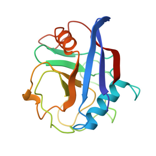
Cyclophilins constitute a family of proteins involved in many essential cellular functions. They have also been identified as a panallergen family able to elicit IgE-mediated hypersensitivity reactions. Moreover, it has been shown that human cyclophilins are recognized by serum IgE from patients sensitized to environmental cyclophilins. IgE-mediated autoreactivity to self-antigens that have similarity to environmental allergens is often observed in atopic disorders. Therefore comparison of the crystal structure of human proteins with similarity to allergens should allow the identification of structural similarities to rationally explain autoreactivity. A new cyclophilin from Aspergillus fumigatus (Asp f 27) has been cloned, expressed and showed to exhibit cross-reactivity in vitro and in vivo. The three-dimensional structure of cyclophilin from the yeast Malassezia sympodialis (Mala s 6) has been determined at 1.5 A (1 A=0.1 nm) by X-ray diffraction. Crystals belong to space group P4(1)2(1)2 with unit cell dimensions of a=b=71.99 A and c=106.18 A. The structure was solved by molecular replacement using the structure of human cyclophilin A as the search model. The refined structure includes all 162 amino acids of Mala s 6, an active-site-bound Ala-Pro dipeptide and 173 water molecules, with a crystallographic R- and free R-factor of 14.3% and 14.9% respectively. The overall structure consists of an eight-stranded antiparallel beta-barrel and two alpha-helices covering the top and bottom of the barrel, typical for cyclophilins. We identified conserved solvent-exposed residues in the fungal and human structures that are potentially involved in the IgE-mediated cross-reactivity.
Organizational Affiliation:
Swiss Institute of Allergy and Asthma Research (SIAF), CH-7270 Davos, Switzerland.