2C9M

Structure of (SR) Calcium-ATPase in the Ca2E1 state solved in a P1 crystal form.


Experimental Data Snapshot

  • Method: X-RAY DIFFRACTION
  • Resolution: 3.00 Å
  • R-Value Free: 0.317 
  • R-Value Work: 0.266 
  • R-Value Observed: 0.266 

wwPDB Validation   3D Report Full Report


This is version 1.3 of the entry. See complete history


Literature

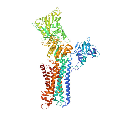
Modulatory and Catalytic Modes of ATP Binding by the Calcium Pump

Lund Jensen, A.-M.Sorensen, T.L.-M.Olesen, C.Moller, J.V.Nissen, P.

(2006) EMBO J 25: 2305

  • DOI: https://doi.org/10.1038/sj.emboj.7601135
  • Primary Citation of Related Structures:  
    2C88, 2C8K, 2C8L, 2C9M

  • PubMed Abstract: 

    We present crystal structures of the calcium-free E2 state of the sarcoplasmic reticulum Ca2+ -ATPase, stabilized by the inhibitor thapsigargin and the ATP analog AMPPCP. The structures allow us to describe the ATP binding site in a modulatory mode uncoupled from the Asp351 phosphorylation site. The Glu439 side chain interacts with AMPPCP via an Mg2+ ion in accordance with previous Fe2+ -cleavage studies implicating this residue in the ATPase cycle and in magnesium binding. Functional data on Ca2+ mediated activation indicate that the crystallized state represents an initial stage of ATP modulated deprotonation of E2, preceding the binding of Ca2+ ions in the membrane from the cytoplasmic side. We propose a mechanism of Ca2+ activation of phosphorylation leading directly from the compact E2-ATP form to the Ca2E1-ATP state. In addition, a role of Glu439 in ATP modulation of other steps of the functional cycle is suggested.


  • Organizational Affiliation

    Department of Molecular Biology, Aarhus University, Denmark.


Macromolecules
Find similar proteins by:  (by identity cutoff)  |  3D Structure
Entity ID: 1
MoleculeChains Sequence LengthOrganismDetailsImage
SARCOPLASMIC/ENDOPLASMIC RETICULUM CALCIUM ATPASE 1
A, B
994Oryctolagus cuniculusMutation(s): 0 
EC: 3.6.3.8
Membrane Entity: Yes 
UniProt
Find proteins for P04191 (Oryctolagus cuniculus)
Explore P04191 
Go to UniProtKB:  P04191
Entity Groups  
Sequence Clusters30% Identity50% Identity70% Identity90% Identity95% Identity100% Identity
UniProt GroupP04191
Sequence Annotations
Expand
  • Reference Sequence
Small Molecules
Ligands 3 Unique
IDChains Name / Formula / InChI Key2D Diagram3D Interactions
CA
Query on CA

Download Ideal Coordinates CCD File 
C [auth A]
D [auth A]
F [auth A]
G [auth A]
I [auth B]
C [auth A],
D [auth A],
F [auth A],
G [auth A],
I [auth B],
J [auth B],
L [auth B]
CALCIUM ION
Ca
BHPQYMZQTOCNFJ-UHFFFAOYSA-N
K
Query on K

Download Ideal Coordinates CCD File 
E [auth A],
K [auth B]
POTASSIUM ION
K
NPYPAHLBTDXSSS-UHFFFAOYSA-N
CL
Query on CL

Download Ideal Coordinates CCD File 
H [auth A],
M [auth B]
CHLORIDE ION
Cl
VEXZGXHMUGYJMC-UHFFFAOYSA-M
Experimental Data & Validation

Experimental Data

  • Method: X-RAY DIFFRACTION
  • Resolution: 3.00 Å
  • R-Value Free: 0.317 
  • R-Value Work: 0.266 
  • R-Value Observed: 0.266 
  • Space Group: P 1
Unit Cell:
Length ( Å )Angle ( ˚ )
a = 64.948α = 97.64
b = 81.276β = 99.94
c = 131.006γ = 95.22
Software Package:
Software NamePurpose
CNSrefinement
DENZOdata reduction
SCALEPACKdata scaling
PHASERphasing

Structure Validation

View Full Validation Report


Entry History 

Deposition Data

Revision History  (Full details and data files)

  • Version 1.0: 2006-12-18
    Type: Initial release
  • Version 1.1: 2011-05-08
    Changes: Version format compliance
  • Version 1.2: 2011-07-13
    Changes: Version format compliance
  • Version 1.3: 2023-12-13
    Changes: Data collection, Database references, Derived calculations, Other, Refinement description