Mechanistic Implications of the Structure of the Mixed-Disulfide Intermediate of the Disulfide Oxidoreductase, 2-Ketopropyl-Coenzyme M Oxidoreductase/Carboxylase.
Pandey, A.S., Nocek, B., Clark, D.D., Ensign, S.A., Peters, J.W.(2006) Biochemistry 45: 113
- PubMed: 16388586
- DOI: https://doi.org/10.1021/bi051518o
- Primary Citation of Related Structures:
2C3C, 2C3D - PubMed Abstract:
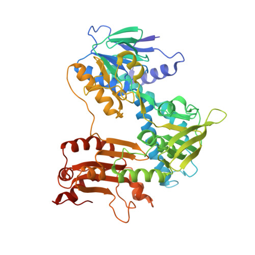
The structure of the mixed, enzyme-cofactor disulfide intermediate of ketopropyl-coenzyme M oxidoreductase/carboxylase has been determined by X-ray diffraction methods. Ketopropyl-coenzyme M oxidoreductase/carboxylase belongs to a family of pyridine nucleotide-containing flavin-dependent disulfide oxidoreductases, which couple the transfer of hydride derived from the NADPH to the reduction of protein cysteine disulfide. Ketopropyl-coenzyme M oxidoreductase/carboxylase, a unique member of this enzyme class, catalyzes thioether bond cleavage of the substrate, 2-ketopropyl-coenzyme M, and carboxylation of what is thought to be an enzyme-stabilized enolacetone intermediate. The mixed disulfide of 2-ketopropyl-coenzyme M oxidoreductase/carboxylase was captured through crystallization of the enzyme with the physiological products of the reaction, acetoacetate, coenzyme M, and NADP, and reduction of the crystals with dithiothreitol just prior to data collection. Density in the active-site environment consistent with acetone, the product of reductive decarboxylation of acetoacetate, was revealed in this structure in addition to a well-defined hydrophobic pocket or channel that could be involved in the access for carbon dioxide. The analysis of this structure and that of a coenzyme-M-bound form provides insights into the stabilization of intermediates, substrate carboxylation, and product release.
Organizational Affiliation:
Department of Chemistry and Biochemistry, Montana State University, Bozeman, Montana 59717, USA.