2C2B

Crystallographic structure of Arabidopsis thaliana Threonine synthase complexed with pyridoxal phosphate and S-adenosylmethionine


Experimental Data Snapshot

  • Method: X-RAY DIFFRACTION
  • Resolution: 2.60 Å
  • R-Value Free: 0.248 
  • R-Value Work: 0.206 
  • R-Value Observed: 0.208 

wwPDB Validation 

Currently 2C2B does not have a validation slider image.


This is version 1.3 of the entry. See complete history


Literature

Allosteric Threonine Synthase: Reorganization of the Pyridoxal Phosphate Site Upon Asymmetric Activation Through S-Adenosylmethionine Binding to a Novel Site.

Mas-Droux, C.Biou, V.Dumas, R.

(2006) J Biol Chem 281: 5188

  • DOI: https://doi.org/10.1074/jbc.M509798200
  • Primary Citation of Related Structures:  
    2C2B, 2C2G

  • PubMed Abstract: 

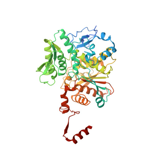
    Threonine synthase (TS) is a fold-type II pyridoxal phosphate (PLP)-dependent enzyme that catalyzes the ultimate step of threonine synthesis in plants and microorganisms. Unlike the enzyme from microorganisms, plant TS is activated by S-adenosylmethionine (AdoMet). The mechanism of activation has remained unknown up to now. We report here the crystallographic structures of Arabidopsis thaliana TS in complex with PLP (aTS) and with PLP and AdoMet (aTS-AdoMet), which show with atomic detail how AdoMet activates TS. The aTS structure reveals a PLP orientation never previously observed for a type II PLP-dependent enzyme and explains the low activity of plant TS in the absence of its allosteric activator. The aTS-AdoMet structure shows that activation of the enzyme upon AdoMet binding triggers a large reorganization of active site loops in one monomer of the structural dimer and allows the displacement of PLP to its active conformation. Comparison with other TS structures shows that activation of the second monomer may be triggered by substrate binding. This structure also discloses a novel fold for two AdoMet binding sites located at the dimer interface, each site containing two AdoMet effectors bound in tandem. Moreover, aTS-AdoMet is the first structure of an enzyme that uses AdoMet as an allosteric effector.


  • Organizational Affiliation

    Laboratoire de Physiologie Cellulaire Végétale, Département Réponse et Dynamique Cellulaires, CNRS Commissariat à l'Energie Atomique, 38054 Grenoble, France.


Macromolecules
Find similar proteins by:  (by identity cutoff)  |  3D Structure
Entity ID: 1
MoleculeChains Sequence LengthOrganismDetailsImage
THREONINE SYNTHASE 1, CHLOROPLASTIC
A, B, C, D, E
A, B, C, D, E, F
486Arabidopsis thalianaMutation(s): 0 
EC: 4.2.3.1
UniProt
Find proteins for Q9S7B5 (Arabidopsis thaliana)
Explore Q9S7B5 
Go to UniProtKB:  Q9S7B5
Entity Groups  
Sequence Clusters30% Identity50% Identity70% Identity90% Identity95% Identity100% Identity
UniProt GroupQ9S7B5
Sequence Annotations
Expand
  • Reference Sequence
Small Molecules
Ligands 3 Unique
IDChains Name / Formula / InChI Key2D Diagram3D Interactions
SAM
Query on SAM

Download Ideal Coordinates CCD File 
G [auth A]
H [auth A]
K [auth B]
L [auth B]
N [auth C]
G [auth A],
H [auth A],
K [auth B],
L [auth B],
N [auth C],
O [auth C],
R [auth D],
S [auth D],
U [auth E],
V [auth E],
X [auth F],
Y [auth F]
S-ADENOSYLMETHIONINE
C15 H22 N6 O5 S
MEFKEPWMEQBLKI-FCKMPRQPSA-N
PLP
Query on PLP

Download Ideal Coordinates CCD File 
AA [auth F]
J [auth A]
M [auth B]
Q [auth C]
T [auth D]
AA [auth F],
J [auth A],
M [auth B],
Q [auth C],
T [auth D],
W [auth E]
PYRIDOXAL-5'-PHOSPHATE
C8 H10 N O6 P
NGVDGCNFYWLIFO-UHFFFAOYSA-N
TRS
Query on TRS

Download Ideal Coordinates CCD File 
I [auth A],
P [auth C],
Z [auth F]
2-AMINO-2-HYDROXYMETHYL-PROPANE-1,3-DIOL
C4 H12 N O3
LENZDBCJOHFCAS-UHFFFAOYSA-O
Experimental Data & Validation

Experimental Data

  • Method: X-RAY DIFFRACTION
  • Resolution: 2.60 Å
  • R-Value Free: 0.248 
  • R-Value Work: 0.206 
  • R-Value Observed: 0.208 
  • Space Group: C 1 2 1
Unit Cell:
Length ( Å )Angle ( ˚ )
a = 191.92α = 90
b = 110.846β = 89.98
c = 152.779γ = 90
Software Package:
Software NamePurpose
REFMACrefinement
MOSFLMdata reduction
SCALAdata scaling
MOLREPphasing

Structure Validation

Currently 2C2B does not have a validation slider image.


Entry History 

Deposition Data

Revision History  (Full details and data files)

  • Version 1.0: 2005-11-28
    Type: Initial release
  • Version 1.1: 2011-07-13
    Changes: Advisory, Refinement description, Version format compliance
  • Version 1.2: 2015-11-11
    Changes: Atomic model, Derived calculations, Non-polymer description, Other, Refinement description, Structure summary
  • Version 1.3: 2023-12-13
    Changes: Data collection, Database references, Derived calculations, Other, Refinement description