2BNX

Crystal structure of the dimeric regulatory domain of mouse diaphaneous-related formin (DRF), mDia1


Experimental Data Snapshot

  • Method: X-RAY DIFFRACTION
  • Resolution: 2.40 Å
  • R-Value Free: 0.236 
  • R-Value Work: 0.196 
  • R-Value Observed: 0.197 

wwPDB Validation   3D Report Full Report


This is version 1.2 of the entry. See complete history


Literature

Structural Basis of Rho Gtpase-Mediated Activation of the Formin Mdia1

Otomo, T.Otomo, C.Tomchick, D.R.Machius, M.Rosen, M.K.

(2005) Mol Cell 18: 273

  • DOI: https://doi.org/10.1016/j.molcel.2005.04.002
  • Primary Citation of Related Structures:  
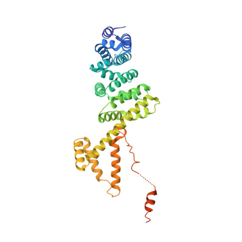
    2BNX

  • PubMed Abstract: 

    Diaphanous-related formins (DRFs) regulate dynamics of unbranched actin filaments during cell contraction and cytokinesis. DRFs are autoinhibited through intramolecular binding of a Diaphanous autoinhibitory domain (DAD) to a conserved N-terminal regulatory element. Autoinhibition is relieved through binding of the GTPase RhoA to the N-terminal element. We report the crystal structure of the dimeric regulatory domain of the DRF, mDia1. Dimerization is mediated by an intertwined six-helix bundle, from which extend two Diaphanous inhibitory domains (DIDs) composed of five armadillo repeats. NMR and biochemical mapping indicate the RhoA and DAD binding sites on the DID partially overlap, explaining activation of mDia1 by the GTPase. RhoA binding also requires an additional structurally independent segment adjacent to the DID. This regulatory construction, involving a GTPase binding site spanning a flexibly tethered arm and the inhibitory module, is observed in many autoinhibited effectors of Ras superfamily GTPases, suggesting evolutionary pressure for this design.


  • Organizational Affiliation

    Department of Biochemistry, University of Texas Southwestern Medical Center at Dallas, 5323 Harry Hines Boulevard, Dallas, Texas 75390, USA.


Macromolecules
Find similar proteins by:  (by identity cutoff)  |  3D Structure
Entity ID: 1
MoleculeChains Sequence LengthOrganismDetailsImage
DIAPHANOUS PROTEIN HOMOLOG 1
A, B
386Mus musculusMutation(s): 0 
UniProt & NIH Common Fund Data Resources
Find proteins for O08808 (Mus musculus)
Explore O08808 
Go to UniProtKB:  O08808
IMPC:  MGI:1194490
Entity Groups  
Sequence Clusters30% Identity50% Identity70% Identity90% Identity95% Identity100% Identity
UniProt GroupO08808
Sequence Annotations
Expand
  • Reference Sequence
Small Molecules
Ligands 1 Unique
IDChains Name / Formula / InChI Key2D Diagram3D Interactions
CL
Query on CL

Download Ideal Coordinates CCD File 
C [auth A]CHLORIDE ION
Cl
VEXZGXHMUGYJMC-UHFFFAOYSA-M
Experimental Data & Validation

Experimental Data

  • Method: X-RAY DIFFRACTION
  • Resolution: 2.40 Å
  • R-Value Free: 0.236 
  • R-Value Work: 0.196 
  • R-Value Observed: 0.197 
  • Space Group: P 32
Unit Cell:
Length ( Å )Angle ( ˚ )
a = 121.365α = 90
b = 121.365β = 90
c = 95.096γ = 120
Software Package:
Software NamePurpose
REFMACrefinement
HKL-2000data reduction
HKL-2000data scaling
CNSphasing

Structure Validation

View Full Validation Report


Entry History 

Deposition Data

Revision History  (Full details and data files)

  • Version 1.0: 2005-06-13
    Type: Initial release
  • Version 1.1: 2011-07-13
    Changes: Refinement description, Version format compliance
  • Version 1.2: 2019-05-08
    Changes: Data collection, Experimental preparation, Other