2BN4

A second FMN-binding site in yeast NADPH-cytochrome P450 reductase suggests a novel mechanism of electron transfer by diflavin reductase


Experimental Data Snapshot

  • Method: X-RAY DIFFRACTION
  • Resolution: 2.91 Å
  • R-Value Free: 0.300 
  • R-Value Work: 0.233 
  • R-Value Observed: 0.233 

wwPDB Validation   3D Report Full Report


Ligand Structure Quality Assessment 


This is version 1.5 of the entry. See complete history


Literature

A Second Fmn-Binding Site in Yeast Nadph-Cytochrome P450 Reductase Suggests a Mechanism of Electron Transfer by Diflavin Reductases.

Lamb, D.C.Kim, Y.Yermalitskaya, L.V.Yermalitsky, V.N.Lepesheva, G.I.Kelly, S.L.Waterman, M.R.Podust, L.M.

(2006) Structure 14: 51

  • DOI: https://doi.org/10.1016/j.str.2005.09.015
  • Primary Citation of Related Structures:  
    2BF4, 2BN4

  • PubMed Abstract: 

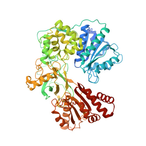
    NADPH-cytochrome P450 reductase transfers two reducing equivalents derived from a hydride ion of NADPH via FAD and FMN to the large family of microsomal cytochrome P450 monooxygenases in one-electron transfer steps. The mechanism of electron transfer by diflavin reductases remains elusive and controversial. Here, we determined the crystal structure of truncated yeast NADPH-cytochrome P450 reductase, which is functionally active toward its physiological substrate cytochrome P450, and discovered a second FMN binding site at the interface of the connecting and FMN binding domains. The two FMN binding sites have different accessibilities to the bulk solvent and different amino acid environments, suggesting stabilization of different electronic structures of the reduced flavin. Since only one FMN cofactor is required for function, a hypothetical mechanism of electron transfer is discussed that proposes shuttling of a single FMN between these two sites coupled with the transition between two semiquinone forms, neutral (blue) and anionic (red).


  • Organizational Affiliation

    Wolfson Laboratory of P450 Biodiversity, Swansea Medical School University of Wales Swansea, Swansea, Wales SA2 8PP, UK.


Macromolecules
Find similar proteins by:  (by identity cutoff)  |  3D Structure
Entity ID: 1
MoleculeChains Sequence LengthOrganismDetailsImage
NADPH CYTOCHROME P450 REDUCTASE
A, B
682Saccharomyces cerevisiaeMutation(s): 0 
EC: 1.6.2.4
UniProt
Find proteins for P16603 (Saccharomyces cerevisiae (strain ATCC 204508 / S288c))
Explore P16603 
Go to UniProtKB:  P16603
Entity Groups  
Sequence Clusters30% Identity50% Identity70% Identity90% Identity95% Identity100% Identity
UniProt GroupP16603
Sequence Annotations
Expand
  • Reference Sequence
Experimental Data & Validation

Experimental Data

  • Method: X-RAY DIFFRACTION
  • Resolution: 2.91 Å
  • R-Value Free: 0.300 
  • R-Value Work: 0.233 
  • R-Value Observed: 0.233 
  • Space Group: P 21 21 21
Unit Cell:
Length ( Å )Angle ( ˚ )
a = 78.14α = 90
b = 77.843β = 90
c = 261.495γ = 90
Software Package:
Software NamePurpose
CNSrefinement
HKL-2000data reduction
SCALEPACKdata scaling
CNSphasing

Structure Validation

View Full Validation Report


Ligand Structure Quality Assessment 


Entry History 

Deposition Data

Revision History  (Full details and data files)

  • Version 1.0: 2006-01-17
    Type: Initial release
  • Version 1.1: 2011-05-08
    Changes: Version format compliance
  • Version 1.2: 2011-07-13
    Changes: Version format compliance
  • Version 1.3: 2019-05-08
    Changes: Data collection, Experimental preparation, Other
  • Version 1.4: 2019-11-06
    Changes: Data collection, Other
  • Version 1.5: 2023-12-13
    Changes: Data collection, Database references, Derived calculations, Refinement description