Structure and Mechanism of the Alkyl Hydroperoxidase Ahpc, a Key Element of the Mycobacterium Tuberculosis Defense System Against Oxidative Stress.
Guimaraes, B.G., Souchon, H., Honore, N., Saint-Joanis, B., Brosch, R., Shepard, W., Cole, S.T., Alzari, P.M.(2005) J Biol Chem 280: 25735
- PubMed: 15886207
- DOI: https://doi.org/10.1074/jbc.M503076200
- Primary Citation of Related Structures:
2BMX - PubMed Abstract:
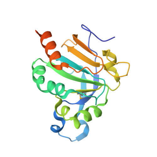
The peroxiredoxin AhpC from Mycobacterium tuberculosis (MtAhpC) is the foremost element of a NADH-dependent peroxidase and peroxynitrite reductase system, where it directly reduces peroxides and peroxynitrite and is in turn reduced by AhpD and other proteins. Overexpression of MtAhpC in isoniazid-resistant strains of M. tuberculosis harboring mutations in the catalase/peroxidase katG gene provides antioxidant protection and may substitute for the lost enzyme activities. We report here the crystal structure of oxidized MtAhpC trapped in an intermediate oligomeric state of its catalytic cycle. The overall structure folds into a ring-shaped hexamer of dimers instead of the usual pentamer of dimers observed in other reduced peroxiredoxins. Although the general structure of the functional dimer is similar to that of other 2-Cys peroxiredoxins, the alpha-helix containing the peroxidatic cysteine Cys61 undergoes a unique rigid-body movement to allow the formation of the disulfide bridge with the resolving cysteine Cys174. This conformational rearrangement creates a large internal cavity enclosing the active site, which might be exploited for the design of inhibitors that could block the catalytic cycle. Structural and mutagenesis evidence points to a model for the electron transfer pathway in MtAhpC that accounts for the unusual involvement of three cysteine residues in catalysis and suggests a mechanism by which MtAhpC can specifically interact with different redox partners.
Organizational Affiliation:
Unité de Biochimie Structurale, CNRS URA 2185, 25 rue du Docteur Roux and Unité de Génétique Moléculaire Bactérienne, Institut Pasteur, 28 rue du Docteur Roux, 75724 Paris.