2BIF

6-PHOSPHOFRUCTO-2-KINASE/FRUCTOSE-2,6-BISPHOSPHATASE H256A MUTANT WITH F6P IN PHOSPHATASE ACTIVE SITE


Experimental Data Snapshot

  • Method: X-RAY DIFFRACTION
  • Resolution: 2.40 Å
  • R-Value Free: 0.244 
  • R-Value Work: 0.200 
  • R-Value Observed: 0.200 

wwPDB Validation   3D Report Full Report


Ligand Structure Quality Assessment 


This is version 1.5 of the entry. See complete history


Literature

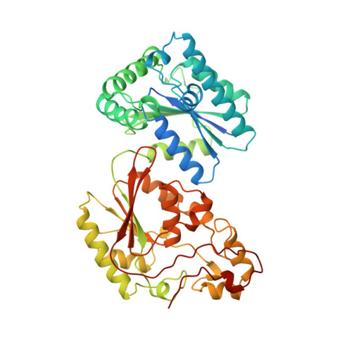
Crystal structure of the H256A mutant of rat testis fructose-6-phosphate,2-kinase/fructose-2,6-bisphosphatase. Fructose 6-phosphate in the active site leads to mechanisms for both mutant and wild type bisphosphatase activities.

Yuen, M.H.Mizuguchi, H.Lee, Y.H.Cook, P.F.Uyeda, K.Hasemann, C.A.

(1999) J Biol Chem 274: 2176-2184

  • DOI: https://doi.org/10.1074/jbc.274.4.2176
  • Primary Citation of Related Structures:  
    2BIF

  • PubMed Abstract: 

    Fructose-6-phosphate,2-kinase/fructose-2,6-bisphosphatase (Fru-6-P, 2-kinase/Fru-2,6-Pase) is a bifunctional enzyme, catalyzing the interconversion of beta-D-fructose- 6-phosphate (Fru-6-P) and fructose-2,6-bisphosphate (Fru-2,6-P2) at distinct active sites. A mutant rat testis isozyme with an alanine replacement for the catalytic histidine (H256A) in the Fru-2,6-Pase domain retains 17% of the wild type activity (Mizuguchi, H., Cook, P. F., Tai, C-H., Hasemann, C. A., and Uyeda, K. (1998) J. Biol. Chem. 274, 2166-2175). We have solved the crystal structure of H256A to a resolution of 2. 4 A by molecular replacement. Clear electron density for Fru-6-P is found at the Fru-2,6-Pase active site, revealing the important interactions in substrate/product binding. A superposition of the H256A structure with the RT2K-Wo structure reveals no significant reorganization of the active site resulting from the binding of Fru-6-P or the H256A mutation. Using this superposition, we have built a view of the Fru-2,6-P2-bound enzyme and identify the residues responsible for catalysis. This analysis yields distinct catalytic mechanisms for the wild type and mutant proteins. The wild type mechanism would lead to an inefficient transfer of a proton to the leaving group Fru-6-P, which is consistent with a view of this event being rate-limiting, explaining the extremely slow turnover (0. 032 s-1) of the Fru-2,6-Pase in all Fru-6-P,2-kinase/Fru-2,6-Pase isozymes.


  • Organizational Affiliation

    Department of Internal Medicine, University of Texas Southwestern Medical Center, Dallas, Texas 75235, USA.


Macromolecules
Find similar proteins by:  (by identity cutoff)  |  3D Structure
Entity ID: 1
MoleculeChains Sequence LengthOrganismDetailsImage
PROTEIN (6-PHOSPHOFRUCTO-2-KINASE/FRUCTOSE-2,6-BISPHOSPHATASE)
A, B
469Rattus norvegicusMutation(s): 5 
Gene Names: RT2K
EC: 2.7.1.105 (PDB Primary Data), 3.1.3.46 (PDB Primary Data)
UniProt
Find proteins for P25114 (Rattus norvegicus)
Explore P25114 
Go to UniProtKB:  P25114
Entity Groups  
Sequence Clusters30% Identity50% Identity70% Identity90% Identity95% Identity100% Identity
UniProt GroupP25114
Sequence Annotations
Expand
  • Reference Sequence
Small Molecules
Ligands 6 Unique
IDChains Name / Formula / InChI Key2D Diagram3D Interactions
ANP
Query on ANP

Download Ideal Coordinates CCD File 
H [auth A]PHOSPHOAMINOPHOSPHONIC ACID-ADENYLATE ESTER
C10 H17 N6 O12 P3
PVKSNHVPLWYQGJ-KQYNXXCUSA-N
BOG
Query on BOG

Download Ideal Coordinates CCD File 
C [auth A],
E [auth A],
J [auth B]
octyl beta-D-glucopyranoside
C14 H28 O6
HEGSGKPQLMEBJL-RKQHYHRCSA-N
F6P
Query on F6P

Download Ideal Coordinates CCD File 
D [auth A],
K [auth B]
6-O-phosphono-beta-D-fructofuranose
C6 H13 O9 P
BGWGXPAPYGQALX-ARQDHWQXSA-N
SIN
Query on SIN

Download Ideal Coordinates CCD File 
I [auth A],
N [auth B]
SUCCINIC ACID
C4 H6 O4
KDYFGRWQOYBRFD-UHFFFAOYSA-N
PO4
Query on PO4

Download Ideal Coordinates CCD File 
G [auth A],
L [auth B],
M [auth B]
PHOSPHATE ION
O4 P
NBIIXXVUZAFLBC-UHFFFAOYSA-K
MG
Query on MG

Download Ideal Coordinates CCD File 
F [auth A]MAGNESIUM ION
Mg
JLVVSXFLKOJNIY-UHFFFAOYSA-N
Experimental Data & Validation

Experimental Data

  • Method: X-RAY DIFFRACTION
  • Resolution: 2.40 Å
  • R-Value Free: 0.244 
  • R-Value Work: 0.200 
  • R-Value Observed: 0.200 
  • Space Group: P 1
Unit Cell:
Length ( Å )Angle ( ˚ )
a = 61.74α = 116.9
b = 73.51β = 99.31
c = 76.7γ = 105.2
Software Package:
Software NamePurpose
X-PLORmodel building
X-PLORrefinement
DENZOdata reduction
SCALEPACKdata scaling
X-PLORphasing

Structure Validation

View Full Validation Report


Ligand Structure Quality Assessment 


Entry History 

Deposition Data

Revision History  (Full details and data files)

  • Version 1.0: 1999-02-16
    Type: Initial release
  • Version 1.1: 2007-10-16
    Changes: Version format compliance
  • Version 1.2: 2011-07-13
    Changes: Version format compliance
  • Version 1.3: 2020-07-29
    Type: Remediation
    Reason: Carbohydrate remediation
    Changes: Data collection, Derived calculations, Structure summary
  • Version 1.4: 2021-11-03
    Changes: Database references, Structure summary
  • Version 1.5: 2023-08-23
    Changes: Data collection, Refinement description