2AVJ

G4(Br)UTTG4 dimeric quadruplex


Experimental Data Snapshot

  • Method: X-RAY DIFFRACTION
  • Resolution: 2.39 Å
  • R-Value Free: 0.242 
  • R-Value Work: 0.168 
  • R-Value Observed: 0.172 

wwPDB Validation   3D Report Full Report


This is version 1.4 of the entry. See complete history


Literature

Topology variation and loop structural homology in crystal and simulated structures of a bimolecular DNA quadruplex.

Hazel, P.Parkinson, G.N.Neidle, S.

(2006) J Am Chem Soc 128: 5480-5487

  • DOI: https://doi.org/10.1021/ja058577+
  • Primary Citation of Related Structures:  
    2AVH, 2AVJ

  • PubMed Abstract: 

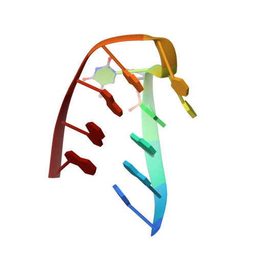
    The topology of DNA quadruplexes depends on the nature and number of the nucleotides linking G-quartet motifs. To assess the effects of a three-nucleotide TTT linker, the crystal structure of the DNA sequence d(G(4)T(3)G(4)) has been determined at 1.5 A resolution, together with that of the brominated analogue d(G(4)(Br)UTTG(4)) at 2.4 A resolution. Both sequences form bimolecular intermolecular G-quadruplexes with lateral loops. d(G(4)(Br)UTTG(4)) crystallized in the monoclinic space group P2(1) with three quadruplex molecules in the asymmetric unit, two associating together as a head-to-head stacked dimer, and the third as a single head-to-tail dimer. The head-to-head dimers have two lateral loops on the same G-quadruplex face and form an eight-G-quartet stack, with a linear array of seven K(+) ions between the quartets. d(G(4)T(3)G(4)) crystallized in the orthorhombic space group C222 and has a structure very similar to the head-to-tail dimer in the P2(1) unit cell. The sequence studied here is able to form several different folds; however, all four quadruplexes in the two structures have lateral loops, in contrast to the diagonal loops reported for the analogous quadruplex with T(4) loops. A total of seven independent T(3) loops were observed in the two structures. These can be classified into two discrete conformational classes, suggesting that these represent preferred loop conformations that are independent of crystal-packing forces.


  • Organizational Affiliation

    Cancer Research U.K. Biomolecular Structure Group, The School of Pharmacy, University of London, 29-39 Brunswick Square, London WC1N 1AX, UK.


Macromolecules

Find similar nucleic acids by:  Sequence   |   3D Structure  

Entity ID: 1
MoleculeChains LengthOrganismImage
5'-D(*GP*GP*GP*GP*(BRU)P*TP*TP*GP*GP*GP*G)-3'
A, B, C, D, E
A, B, C, D, E, F
11N/A
Sequence Annotations
Expand
  • Reference Sequence
Small Molecules
Ligands 2 Unique
IDChains Name / Formula / InChI Key2D Diagram3D Interactions
CAC
Query on CAC

Download Ideal Coordinates CCD File 
P [auth D],
Q [auth D]
CACODYLATE ION
C2 H6 As O2
OGGXGZAMXPVRFZ-UHFFFAOYSA-M
K
Query on K

Download Ideal Coordinates CCD File 
G [auth A]
H [auth A]
I [auth A]
J [auth A]
K [auth C]
G [auth A],
H [auth A],
I [auth A],
J [auth A],
K [auth C],
L [auth C],
M [auth C],
N [auth C],
O [auth C],
R [auth E],
S [auth E],
T [auth E],
U [auth E],
V [auth F]
POTASSIUM ION
K
NPYPAHLBTDXSSS-UHFFFAOYSA-N
Experimental Data & Validation

Experimental Data

  • Method: X-RAY DIFFRACTION
  • Resolution: 2.39 Å
  • R-Value Free: 0.242 
  • R-Value Work: 0.168 
  • R-Value Observed: 0.172 
  • Space Group: P 1 21 1
Unit Cell:
Length ( Å )Angle ( ˚ )
a = 31.681α = 90
b = 33.309β = 91.49
c = 79.234γ = 90
Software Package:
Software NamePurpose
DENZOdata reduction
SCALEPACKdata scaling
SHELXphasing
REFMACrefinement
PDB_EXTRACTdata extraction
SHELXDphasing

Structure Validation

View Full Validation Report


Entry History 

Deposition Data

Revision History  (Full details and data files)

  • Version 1.0: 2006-03-28
    Type: Initial release
  • Version 1.1: 2008-05-01
    Changes: Version format compliance
  • Version 1.2: 2011-07-13
    Changes: Non-polymer description, Version format compliance
  • Version 1.3: 2017-10-11
    Changes: Refinement description
  • Version 1.4: 2024-02-14
    Changes: Data collection, Database references, Derived calculations