2AFT

Formylglycine generating enzyme C336S mutant


Experimental Data Snapshot

  • Method: X-RAY DIFFRACTION
  • Resolution: 1.66 Å
  • R-Value Free: 0.187 
  • R-Value Work: 0.149 
  • R-Value Observed: 0.151 

wwPDB Validation   3D Report Full Report


This is version 2.2 of the entry. See complete history


Literature

A general binding mechanism for all human sulfatases by the formylglycine-generating enzyme

Roeser, D.Preusser-Kunze, A.Schmidt, B.Gasow, K.Wittmann, J.G.Dierks, T.von Figura, K.Rudolph, M.G.

(2006) Proc Natl Acad Sci U S A 103: 81-86

  • DOI: https://doi.org/10.1073/pnas.0507592102
  • Primary Citation of Related Structures:  
    2AFT, 2AFY, 2AII, 2AIJ, 2AIK

  • PubMed Abstract: 

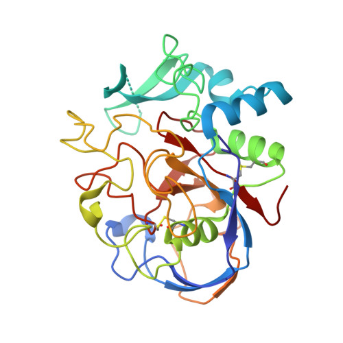
    The formylglycine (FGly)-generating enzyme (FGE) uses molecular oxygen to oxidize a conserved cysteine residue in all eukaryotic sulfatases to the catalytically active FGly. Sulfatases degrade and remodel sulfate esters, and inactivity of FGE results in multiple sulfatase deficiency, a fatal disease. The previously determined FGE crystal structure revealed two crucial cysteine residues in the active site, one of which was thought to be implicated in substrate binding. The other cysteine residue partakes in a novel oxygenase mechanism that does not rely on any cofactors. Here, we present crystal structures of the individual FGE cysteine mutants and employ chemical probing of wild-type FGE, which defined the cysteines to differ strongly in their reactivity. This striking difference in reactivity is explained by the distinct roles of these cysteine residues in the catalytic mechanism. Hitherto, an enzyme-substrate complex as an essential cornerstone for the structural evaluation of the FGly formation mechanism has remained elusive. We also present two FGE-substrate complexes with pentamer and heptamer peptides that mimic sulfatases. The peptides isolate a small cavity that is a likely binding site for molecular oxygen and could host reactive oxygen intermediates during cysteine oxidation. Importantly, these FGE-peptide complexes directly unveil the molecular bases of FGE substrate binding and specificity. Because of the conserved nature of FGE sequences in other organisms, this binding mechanism is of general validity. Furthermore, several disease-causing mutations in both FGE and sulfatases are explained by this binding mechanism.


  • Organizational Affiliation

    Department of Molecular Structural Biology, University of Göttingen, Justus-von-Liebig-Weg 11, D-37077 Göttingen, Germany.


Macromolecules
Find similar proteins by:  (by identity cutoff)  |  3D Structure
Entity ID: 1
MoleculeChains Sequence LengthOrganismDetailsImage
Sulfatase modifying factor 1A [auth X]286Homo sapiensMutation(s): 1 
UniProt & NIH Common Fund Data Resources
Find proteins for Q8NBK3 (Homo sapiens)
Explore Q8NBK3 
Go to UniProtKB:  Q8NBK3
PHAROS:  Q8NBK3
GTEx:  ENSG00000144455 
Entity Groups  
Sequence Clusters30% Identity50% Identity70% Identity90% Identity95% Identity100% Identity
UniProt GroupQ8NBK3
Sequence Annotations
Expand
  • Reference Sequence
Oligosaccharides

Help

Entity ID: 2
MoleculeChains Length2D Diagram Glycosylation3D Interactions
2-acetamido-2-deoxy-beta-D-glucopyranose-(1-4)-2-acetamido-2-deoxy-beta-D-glucopyranoseB [auth A]2N-Glycosylation
Glycosylation Resources
GlyTouCan:  G42666HT
GlyCosmos:  G42666HT
GlyGen:  G42666HT
Experimental Data & Validation

Experimental Data

  • Method: X-RAY DIFFRACTION
  • Resolution: 1.66 Å
  • R-Value Free: 0.187 
  • R-Value Work: 0.149 
  • R-Value Observed: 0.151 
  • Space Group: P 21 21 2
Unit Cell:
Length ( Å )Angle ( ˚ )
a = 61.786α = 90
b = 109.514β = 90
c = 43.425γ = 90
Software Package:
Software NamePurpose
REFMACrefinement
DENZOdata reduction
SCALEPACKdata scaling

Structure Validation

View Full Validation Report


Entry History 

Deposition Data

Revision History  (Full details and data files)

  • Version 1.0: 2005-12-13
    Type: Initial release
  • Version 1.1: 2008-04-30
    Changes: Version format compliance
  • Version 1.2: 2011-07-13
    Changes: Non-polymer description, Version format compliance
  • Version 2.0: 2020-07-29
    Type: Remediation
    Reason: Carbohydrate remediation
    Changes: Atomic model, Data collection, Derived calculations, Structure summary
  • Version 2.1: 2021-11-10
    Changes: Database references, Structure summary
  • Version 2.2: 2023-10-25
    Changes: Data collection, Refinement description