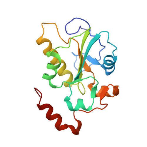
Experimental Validation of the Docking Orientation of Cdc25 with Its Cdk2-CycA Protein Substrate.
Sohn, J., Parks, J.M., Buhrman, G., Brown, P., Safi, A., Edelsbrunner, H., Yang, W., Rudolph, J.(2005) Biochemistry 44: 16563-16573
- PubMed: 16342947
- DOI: https://doi.org/10.1021/bi0516879
- Primary Citation of Related Structures:
2A2K - PubMed Abstract:
Cdc25 phosphatases are key activators of the eukaryotic cell cycle and compelling anticancer targets because their overexpression has been associated with numerous cancers. However, drug discovery targeting these phosphatases has been hampered by the lack of structural information about how Cdc25s interact with their native protein substrates, the cyclin-dependent kinases. Herein, we predict a docked orientation for Cdc25B with its Cdk2-pTpY-CycA protein substrate by a rigid-body docking method and refine the docked models with full-scale molecular dynamics simulations and minimization. We validate the stable ensemble structure experimentally by a variety of in vitro and in vivo techniques. Specifically, we compare our model with a crystal structure of the substrate-trapping mutant of Cdc25B. We identify and validate in vivo a novel hot-spot residue on Cdc25B (Arg492) that plays a central role in protein substrate recognition. We identify a hot-spot residue on the substrate Cdk2 (Asp206) and confirm its interaction with hot-spot residues on Cdc25 using hot-spot swapping and double mutant cycles to derive interaction energies. Our experimentally validated model is consistent with previous studies of Cdk2 and its interaction partners and initiates the opportunity for drug discovery of inhibitors that target the remote binding sites of this protein-protein interaction.
Organizational Affiliation:
Department of Biochemistry, Duke University Medical Center, Durham, North Carolina 27710, USA.